BIOLOGY FORM ONE NOTES

CHAPTER ONE: NUTRITION IN ANIMALS - Biology Form Two

Chapter One: Nutrition in Animals

Introduction

Animals get their food from plants or other animals. They exhibit a heterotrophic mode of nutrition. In this chapter, you will learn about animal nutrition which include food nutrients. The competences developed will enable you to maintain good eating habits so as to enhance general body health and well-being. This tendency will enable you to avoid the risks of getting nutritional diseases and disorders, as well as other nutritional problems.

Think

The life of animals without nutrients involves nutrient requirement, mode of taking food and its utilisation in the animal body. Animal nutrition also focuses on the dietary nutrient needs of animals. Animals are heterotrophs since they depend on other organisms for their nutritional needs.

Concept of Nutrition in Animals

Task 1.1

Search for information from the library and internet sources on the concept of nutrition in animals. Write short notes on searched information.

Nutrition refers to the study of the relationship between diet, health, and diseases. There are two major types of nutrition based on how organisms obtain their food. These are autotrophic and heterotrophic nutrition Animal nutrition.

Heterotrophic Nutrition

Heterotrophs are organisms that cannot make their own food but rely on assuming other organism or organic matter for energy and nutrients. The term 'hetero' means different and 'trophy' means feeding. Therefore, 'heterotroph' means 'different feeding'. Heterotrophs feed on either different food substances manufactured by other organisms or feed on other organisms directly. Examples of heterotrophs include all animals, fungi, most bacteria, and protocysts. The mode of feeding in which an organism is unable to make its own food but depends on food already made by other organisms is called heterotrophic nutrition. There are various types of heterotrophic nutrition, including holozoic, saprophytic, and symbiotic nutrition.

Holozoic Nutrition

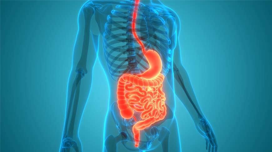
This mode of nutrition involves taking in complex food substances, digesting, absorbing, and assimilating the nutrients into the organism's body. The undigested and indigestible food remains are finally egested as faeces. This type of nutrition is found in most animals. Holozoic nutrition is divided into three modes of feeding: carnivorous, herbivorous and omnivorous.

  • Carnivorous: This is a mode of feeding in which an animal feeds on other animals. Examples of animals that practise this type of feeding are driver ants, ground beetles, lions, tigers, and leopards. These animals are called carnivores because they eat other animals, usually of different species. In this kind of feeding relationship, animals that hunt other animals are called predators, while those that are hunted are called preys.
  • Herbivorous: This is a mode of feeding in which an organism feeds on plants. Animals that undergo this type of feeding are called herbivores because they eat plants or parts of plants. Examples of plant eaters or herbivores include grasshoppers, cattle, rabbits, goats, antelopes, and giraffes.
  • Omnivorous: This is a mode of feeding in which an organism feeds on both plant and animal food sources. They also feed on other organisms including fungi and algae. They are also known as opportunistic feeders because they feed on a variety of food sources. Examples of omnivorous animals include human beings, bears, chimpanzees, birds, pigs, turtles, lizards, and certain insects such as crickets, ants, and wasps.

Saprophytic Nutrition

This is a mode of feeding in which an organism feeds on decaying organic matter. Such decaying matter may originate from plants, animals, and other decomposing materials. Organisms that obtain their food from dead and decaying organic matter are called saprophytes. In this mode of feeding, a saprophyte releases digestive enzymes externally on the substrate. The released enzymes convert complex organic molecules such as starch into simple molecules such as glucose. The glucose can be easily absorbed by body cells and used for various activities. Digestion, which takes place by the release of enzymes outside the cell, is also called extracellular digestion. An example of a saprophyte is a mushroom growing on a log. The mushroom releases enzymes externally through their root-like structures, called rhizoids. The enzymes cause decomposition and decay of the log from which the mushroom gets its nutrients. Another example of a saprophyte is bread mould that grows on the surface of decaying bread to obtain the nutrients.

">

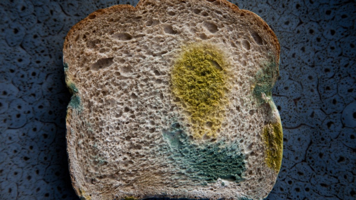
Figure 1.1: Bread mould growing on decaying bread

Symbiotic Nutrition

This is a feeding relationship or an association between two species in which one or both benefit. Symbiosis can be in the form of commensalism, mutualism, or parasitism.

  • Commensalism: This is a feeding relationship or an association between individuals of two different species in which one species benefits, while the other is neither harmed nor benefitted. In most cases, the host individual is larger than the commensal individual. Commensal organisms benefit in various ways such as in getting nutrients, shelter, support, or transport from the host. For example, small fishes like remora attach to the body of a large fish such as a shark. The shark enables remora to move in different areas without using its own energy, but the shark is not harmed, and it does not benefit from such a relationship. Other examples are epiphytes which are small plants that grow on different trees but do not harm the trees. The epiphytes get access to nutrients, exposure to sunlight, and support from the trees while the trees are neither harmed nor benefitted by the relationship.
  • Figure 1.2: Host and epiphytic plants exhibiting commensalism

  • Mutualism: This is a symbiotic relationship between two species in which both species are mutually benefitted. For example, bacteria living in the gut of a goat, sheep, or cow get their nutrients from the food digested by the animals. The bacteria in turn help the animals to digest cellulose, by releasing cellulase enzymes that digest cellulose.
  • Parasitism: This is a kind of feeding relationship between two species where one species benefits while the other is harmed. For example, the relationship between a tapeworm and a human being. The tapeworm lives in the human gut and gets nutrients from the human body while the human being is harmed by getting sick. The organism that benefits is called a parasite while the one that is harmed is called a host. In the human-tapeworm relationship, the human being is a host and the tapeworm is a parasite.

In parasitic mode of nutrition, the parasites depend on nutrients that are found in the body of their hosts. There are two types of parasites, namely ectoparasites and endoparasites. Ectoparasites live outside the body of the host. Examples of ectoparasites include ticks, lice, fleas, and bedbugs. Endoparasites live and obtain their food inside the body of the host. Plasmodium sp., Ascaris sp., and tapeworms are examples of endoparasites.

The Importance of Nutrition

Nutrition is important in various ways. It encompasses the study of the relationship between diet, health, and diseases. People who study nutrition are called nutritionists or dieticians. They are professionals who advise people on how nutrition affects their health and well-being. Through nutrition, an organism can get the necessary nutrients for the body to produce energy and support functioning of various life processes. Such life processes include growth and the development of cells, tissues and organs, the repair of damaged parts, and movement. Other processes include ensuring protection against infections and diseases, maintaining body temperature, and reproduction. Through nutrition the body can also synthesise important chemical substances, such as enzymes and hormones.

Moreover, maintaining good nutrition helps to promote general body health so as to reduce the risk of getting chronic diseases such as heart attack, diabetes, stroke, blood pressure, osteoporosis, and certain types of cancer.

Exercise 1.1

  1. Using relevant examples, briefly explain the following types of heterotrophic nutrition:
    1. holozoic nutrition;
    2. saprophytic nutrition; and
    3. symbiotic nutrition.
  2. Why are omnivores regarded as opportunistic feeders?
  3. What is the role of enzymes in the saprophytic mode of feeding?
  4. Why are large trees in the forest not harmed by the epiphytes that grow on their bodies?
  5. Explain the meaning of the following:
    1. commensalism;
    2. parasitism; and
    3. mutualism.
  6. How would you apply the concept of nutrition to improve your diet?
  7. What would happen to the heterotrophs if there were no autotrophs?
  8. Why is proper nutrition essential for maintaining health.
  9. Why are human beings, pigs, and bears regarded as omnivorous animals?

Nutrients

Task 1.2

Search for information from the library and internet sources on the concept of nutrients. Summarise important points from what you have searched.

Nutrients are chemical components of foods, which are essential to life and health. They are important for promoting growth and development, as well as regulating chemical processes in the body. Nutrients are obtained from various foods such as cassava, meat, bananas and vegetables.

There are five major types of nutrients, which are carbohydrates, proteins, lipids (fats and oil), vitamins, and minerals. Water and roughage are also important. Nutrients can be divided into two categories depending on the quantity needed by the body. These groups are macronutrients and micronutrients. Macronutrients refer to nutrients that are needed by the body in large amounts. They include proteins, carbohydrates, and lipids. These nutrients contain kilocalories (kcal) which can be burned in the body to produce energy needed for various body functions. A calorie is a unit of measurement of food energy. Micronutrients are those needed by the body in small amounts but are essential for carrying out body functions. They usually assist in many aspects of body functions such as producing energy, digesting food substances, and making macromolecules. Examples of micronutrients are vitamins and minerals. Unlike macronutrients, micronutrients do not contain calories.

Activity 1.1: Classify food items according to the nutrients they provide.

Materials: A variety of food items

Procedure:

  1. Collect a variety of food items such as eggs, maize, beans, carrots, potatoes, sunflower seeds, groundnuts, vegetables and tomatoes.
  2. Group such food items into different categories based on the type of nutrients they contain.

Question: How many food items did you manage to classify?

Proteins

Proteins are organic compounds made up of one or more chains of amino acids. Amino acids are made of smaller units that contain carbon, hydrogen, oxygen, nitrogen and sulphur. There are two groups of amino acids: essential amino acids and non-essential amino acids. The essential amino acids are obtained from foods eaten, while non-essential amino acids can be made by the body. However, the body cannot produce essential amino acids. Foods like beef, chicken, fish, termites, eggs, beans, lentils, peas, groundnuts, mushrooms, and milk and its products like cheese are rich in proteins hence provide essential amino acids.

>

Figure 1.3: Sources of protein

Functions of Proteins

Proteins have several functions in the human body including building body cells, promoting growth, and making protoplasm. Proteins also help to repair damaged tissues such as replacing damaged or lost cells in wounds, skin, and mucus membranes. Since proteins are major components of all cell membranes, they play a role in the transport of other molecules across the cell membranes. They form part of the cell structure, and aid in the contraction of muscles. They are sometimes used as a source of energy, especially when the body lacks carbohydrates and lipids. Each gram of protein provides four kilocalories (kcal) of energy. However, proteins are not the major source of energy in the body. Their primary function is to promote growth and repair worn out body tissues. Proteins are essential for growing children, pregnant women, and people recovering from illnesses.

When proteins are consumed, they are broken down into amino acids that are absorbed into the bloodstream and used by the body to produce its own proteins. For example, the haemoglobin found in the red blood cells is one of the body proteins. Haemoglobin enables red blood cells to transport oxygen in our bodies. Other examples of body proteins include antibodies which provide body immunity and enzymes that are involved in food digestion. In addition, many hormones are protein in nature. They control and regulate various processes in the body.

Lipids

They are organic compounds made of carbon, hydrogen, and oxygen. They are insoluble in water but soluble in organic solvents such as ether and benzene. The main forms of dietary lipids are fats and oils. Fats are solid at room temperature while oils are liquid at room temperature. Lipids are made up of fatty acids and glycerol. Fatty acids can be essential or non-essential. Essential fatty acids are obtained from foods eaten while non-essential fatty acids can be made by the body. The body can not produce essential fatty acids. It is, therefore, important to eat foods that contain essential fatty acids. Examples of such foods are oily fish, nuts, avocados, oil, and seeds such as sunflower and sesame.

Figure 1.4: Sources of lipids

Functions of Lipids

Lipids are a source of energy. They provide more energy than all other food substances. Each gram of lipid provides nine kilocalories (kcal) of energy. They are important components of cell membranes. Fat deposits surround and protect body organs such as the heart and kidneys. Stores of fat in the adipose tissue under the skin help to regulate body temperature by insulating the body against loss of heat. Fat-soluble vitamins are also stored in fatty tissues. Essential fatty acids are important for the formation of substances that help to control blood pressure and activate the body's immune response.

Carbohydrates

Carbohydrates are organic compounds made up of small repeating units called monosaccharides. The small repeating units form bond with each other to make a larger molecule. Monosaccharides are mainly made up of carbon, hydrogen, and oxygen. Carbohydrates are sugar molecules found in certain foods and drinks. Sources of carbohydrates include maize, sweet potatoes, sugarcane, bananas, cassava, fruits, and yams.

Figure 1.5: Sources of carbohydrates

Functions of Carbohydrates

Carbohydrates are a major source of energy in the body. In an ideal situation, we should get two-thirds of our energy from carbohydrates. They are broken down into glucose which acts as fuel for the body. Each gram of carbohydrate produces four kilocalories (kcal) of energy in the body to be used by the cells to perform work. Carbohydrates are also essential for the proper functioning of the nervous system, heart, and kidneys. In addition, carbohydrates form genetic materials, which are DNA (Deoxyribonucleic Acid) and RNA (Ribonucleic Acid) in the cells.

Excess carbohydrates in the body are converted into either glycogen or fats in the liver. The glycogen is stored in the liver and muscles, while fats are stored as fat deposits in the lower abdomen, around kidneys, and under the skin. Glycogen acts as a source of energy when there is short supply of carbohydrates in the body. In the muscles and liver, glycogen is broken down to glucose for provision of energy when required, for example, during a vigorous physical activity. Also, carbohydrates combine with proteins and lipids to form glycoproteins and glycolipids, respectively, which are important components of cell membranes.

Vitamins

Vitamins are complex organic micronutrients that are essential for body growth and survival. Animals need vitamins for proper body functioning and metabolism. Vitamins can be grouped into two categories, which are water-soluble and fat-soluble vitamins.

Fat-soluble vitamins can be stored in the body. Vitamins A, D, E and K are examples of fat-soluble vitamins.

Water-soluble vitamins are not stored in the body. Vitamins B and C are water-soluble. Vitamin B is of various forms namely, vitamin B1, B2, B3, B6, B9 and B12. These can be easily destroyed by heat.

Vitamins are formed by the combination of glucose and some mineral elements. Most of the vitamins are synthesised by autotrophs, particularly green plants. Mammals can synthesise vitamins A and D using plant molecules obtained in their food. Examples of foods containing vitamins are fruits and vegetables.

Figure 1.6: Sources of vitamins

Functions of Vitamins

Vitamins play an important role in the metabolic activities of both plants and animals, such as promoting functions of immune enzymes and nervous systems. They are also important in the formation of bone tissue and red blood cells. Deficiency or excess intake of vitamins can cause health problems.

Table 1.1: Sources, functions, and deficiency symptoms of vitamins
Vitamin Sources Functions Deficiency Symptoms
Vitamin A (Retinol) Liver, milk, eggs, oranges, and yellow vegetables such as carrots and pumpkins Formation of membranes of the eyes, and the respiratory tract
  1. Night blindness
  2. Increased risk of infections
Vitamin B1 (Thiamine) Lean meat, liver, eggs, mushroom, brown rice, and whole grain cereals
  1. Assists in carbohydrate metabolism
  2. Helps in muscle coordination
Beriberi, a disease characterised by a loss of appetite, muscle cramps, nerve disorder, and heart failure
Vitamin B2 (Riboflavin) Liver, meat, mushroom, and whole grain cereals Metabolism of all foods, and the release of energy to cells
  1. Cracks and sores around the mouth and nose
  2. Visual problems
Vitamin B3 (Niacin) Nuts, fish, meat, mushroom, brown rice, and whole grain cereals Needed by many enzymes that convert food to energy Pellagra, a disease characterised by skin lesions, loss of appetite, diarrhoea, and muscle weakness
Vitamin B6 (Pyridoxine) Meat, vegetables, mushroom, and whole grain cereals Protein metabolism
  1. Nerve irritability
  2. Sores in the mouth and eyes
  3. Anaemia
Vitamin B9 (Folic acid) Liver, fish, green leafy vegetables, legumes, and folate fortified foods Synthesis of new cells
  1. Birth defects
  2. Anaemia
  3. Frequent infections
Vitamin B12 (Cyanocobalamin) Fish, meat, eggs, milk, and liver
  1. Helps in making genetic material
  2. Formation of red blood cells
  1. Anaemia
  2. Nerve damage
  3. Weight loss
Vitamin C (Ascorbic acid) Tomatoes, fresh green vegetables, and citrus fruits such as oranges and lemons
  1. Used as Antioxidant
  2. Helps in wound healing
  1. Muscle weakness
  2. Easy bruising
  3. Joint pains
  4. Scurvy (bleeding gums)
  5. Poor healing of wounds
  6. Frequent infections
Vitamin D (Calciferol) Egg yolk, milk, oily fish, sunlight, and liver Helps to build and maintain teeth and bones
  1. Rickets in children
  2. Osteoporosis (soft bones) in adults
Vitamin E (Tocopherol) Corn or sunflower oil, butter, brown rice, and peanuts
  1. Used as Antioxidant
  2. Protects damage of cell membranes
  1. Nerve abnormalities
  2. Infertility
Vitamin K Green vegetables, and liver Needed for normal blood clotting Defective blood coagulation resulting in excessive bleeding in case of injury

Minerals

Minerals are inorganic elements that come from the soil and water and are absorbed and used by living organisms. In animal nutrition, minerals fulfill multiple functions in the physiology of all systems. Different types of minerals are needed for different activities in the body. These minerals are called essential minerals. Examples of essential minerals needed by the body include sodium, chlorine, potassium, calcium, phosphorus, magnesium, sulphur, zinc, iodine, copper, manganese and fluoride.

Function of Minerals

Most minerals are important for enzyme activities. Some minerals are essential for maintaining balance of body fluids, development of bone tissue, synthesis of hormones, activation of enzymes, transmission of nerve impulses, as well as contraction and relaxation of muscles.

Table 1.2: Examples of minerals, their sources, functions and deficiency symptoms
Mineral Sources Functions Deficiency Symptoms
Calcium Milk, yoghurt, cheese, sardines, eggs, and green vegetables
  1. Helps build strong bones and teeth
  2. Promotes nerve transmission
  3. Helps in muscle contraction
  4. Important in clotting blood
  1. Weak bones and bleeding easily
  2. Impaired nerve activity
Phosphorus Meat, milk, fish, eggs, and nuts
  1. Builds bones and teeth
  2. Helps maintain acid-base balance
  3. Aids formation of genetic materials
Poor bone and teeth formation
Potassium Peanuts, bananas, orange juice, meat, and green beans Helps maintain regular fluid balance needed for nerve and muscle function
  1. Poor muscle contraction
  2. Impaired nerve activity
Sodium Table salt, soy sauce, milk, bread, vegetables, and processed foods
  1. Necessary for nerve transmission
  2. Needed for muscle contraction
  3. Necessary for protein making
  4. Needed for proper fluid balance
  1. Muscle cramps
  2. Poor nerve transmission
Chloride Table salt, eggs, milk, meat, sea foods, and whole grain foods Maintenance of water and ionic balance in the body and formation of hydrochloric acid in the stomach Poor digestion of proteins
Magnesium Spinach, pumpkin seeds, sesame seeds, beans, nuts, sea foods, and magnesium
  1. Maintaining a healthy immune system
  2. Making proteins
  3. Contraction of muscles
  4. Transmission of nerve impulses
  5. Strengthening of bones
  1. Muscle weakness
  2. Irregular heartbeat
  3. Weak bones
Iodine Sea foods, dairy products, and iodized table salt Production of thyroid hormone which regulate growth, development, and metabolism
  1. Goitre (enlarged thyroid gland)
  2. Growth retardation
  3. Mental retardation
  4. Still birth
Fluoride Fish, fluorinated water or water naturally containing fluorine, and most varieties of tea such as green tea
  1. Development of teeth and bones
  2. Helps to prevent tooth decay
  1. Poor development of bones and teeth
  2. Tooth decay
Manganese Found in most plant foods, kidneys, liver, tea, coffee, nuts, and fruits
  1. Helps in the formation of bones
  2. Activation of enzymes (Coenzyme)
  1. Nausea
  2. Dizziness
  3. Loss of hearing
  4. Loss of bone mass
Copper Whole grains, meat, drinking water, fish, liver, legumes, nuts, and other seeds
  1. Synthesis of bones and haemoglobin
  1. Bleeding under the skin
  2. Easy rupturing of blood vessels
  3. Bones and joints problems
  4. Anaemia
Iron Liver, lean meat, fish, chicken, egg yolk, legumes, kidney, beans, and green vegetables
  1. Essential for making red pigment in blood (haemoglobin)
  2. Essential for energy production
Anaemia
Zinc Oysters, shrimp, crab, meat, fish, chicken, whole grains, and vegetables
  1. Necessary for a healthy immune system
  2. Controls normal growth and sexual maturation
  3. Activation of enzymes
  4. Helps in wound healing
  5. Helps in taste detection
  6. Promotes protein synthesis and DNA production
  1. Impaired taste
  2. Poor immune response
  3. Skin problems

Roughage

This is dietary fibre that is obtained from the indigestible part of plants. Roughage does not have any nutritional value as it is not digested and absorbed in the body. Sources of dietary fibre include cassava, potatoes, beans, fruits, cabbage, spinach, and whole grain cereals.

Figure 1.7: Sources of roughage

Functions of Roughage

Roughage helps in the passage of food and faeces through the gut by aiding contraction and relaxation of the gut muscles (peristalitic movement). It also helps to control body weight, thus protects the body from diseases such as heart diseases.

Water

Water is an essential nutrient for the human body as it is a major constituent of body fluids such as blood, tears, and saliva. It forms a large part of the cell cytoplasm and helps to maintain the shape of cells, tissues, and organs. Water helps in the process of breaking down food during digestion. It also lubricates moving parts of the body and prevents friction. Additionally, water helps in the regulation of body temperature. Sweating is one of the means by which water prevents the human body from overheating during hot weather. This is because evaporation of sweat from the surface of the skin helps the body to cool during hot conditions. It is important to drink enough safe water, and eat raw vegetables and fruits to maintain an optimal water content in the body. A lacking of adequate water in the body leads to thirst, dehydration and constipation. Losing more than 10% of water in the body can lead to death.

Balanced Diet

Task 1.3

Search for information from the library and internet sources on concept of a balanced diet. Then, summarise your findings.

A balanced diet refers to a meal containing foods from all food groups in the right proportions. A proper diet helps an individual to remain healthy. For young individuals, a balanced diet helps them to grow and develop normally. It also help them to maintain an ideal body weight. Moreover, it minimises the risk of contracting long-lasting diseases like cancer, diabetes, obesity, high blood pressure and others.

Components of a balanced diet include:

  1. Carbohydrates (about 60% of daily diet);
  2. Proteins (10-35% according to the physiological needs);
  3. Fats (about 15% of daily calorie needs);
  4. Vitamins and minerals; and
  5. Water

Factors for a Lack of a Balanced Diet

A lack of balanced diet comes from various factors, including the following:

  1. Limited access to nutritious food: People living in food desert or areas with inadequate availability of fresh produce many struggle to get a balanced diet
  2. Economic constraints: Financial limitations can hinder the ability to purchase a variety of healthy foods, leading to an imbalanced diet
  3. Poor dietary choices: High consumption of highly processed food and convenience foods often in a diet that is low in essential nutrients
  4. Lack of knowledge: Insufficient understanding of dietary guidelines can lead to unbalanced food choices. Misinformation about healthy eating can also contribute to poor dietary choices.
  5. Cultural and social influences: Dietary habits influenced by cultural preferences or social norms may not always include a variety of nutrient - dense foods
  6. Time constraints: Busy lifestyles and competing responsibilities can limit the time available for meal planning and preparation, leading to reliance on less nutritious options.
  7. Health condition: Dietary restrictions due to health conditions or allergies can complicate maintaining a balanced diet if suitable alternatives are not included.
  8. Psychological factors: Emotional eating or eating disorders can disrupt balanced eating patterns and lead to unhealthy food choices.

To promote a healthy lifestyle, it is important to take in diets containing low fats, sugars and salts but having moderate amounts of proteins, carbohydrates, vitamins, minerals and roughages. It is also important to drink adequate amounts of water and engage in regular physical exercise.

The following should be done to ensure a healthy lifestyle:

  1. Take recommended amounts of proteins, fats or oils, sugars, and salts;
  2. Take recommended amounts of fruits and vegetables in the diet;
  3. Eat a variety of foods, including whole grains, fruits, vegetables, and food with roughage to provide the body with the required energy and nutrients;
  4. Maintain an appropriate body weight to height ratio within the limits recommended by the World Health Organisation (WHO);
  5. Do physical activities daily for 30 to 40 minutes;
  6. Replace red meat with fish, poultry, or lean meat in your diet;
  7. Take low fat dairy products and low salt foods;
  8. Limit consumption of sugary drinks and all sweets; and
  9. Avoid processed and baked foods, particularly those high in sugars, unhealthy fats and additives.

Nutritional Requirements for Different Groups of People

Task 1.4

Search for information from the library and internet sources on diet requirements for different groups of people. Then, summarise your findings.

The ratio of nutrients needed by the body varies depending on the age, sex, health state, amount of activity, or general state of the body. Therefore, different groups of people such as pregnant women and lactating mothers, children, the elderly, and the sick have different nutritional needs.

Pregnant Women and Lactating Mothers

Pregnant women and lactating mothers need to get enough nutrients in their meals. They should eat a balanced diet because they require enough nutrients for themselves and their growing foetus and babies, respectively. Food rich in protein is needed by pregnant women and lactating mothers to build the muscles, breasts, uterus, blood supply, and for proper functioning of tissues and organs of the foetus and baby.

Pregnant women are advised to eat food rich in folic acid. These help to lower the risk of birth defects such as spina bifida. Spina bifida is a spinal disorder characterised by a hole in the spine. It results from incomplete formation of the spinal cord and the bones of the spine. Often, the spine protrudes through the hole and sometimes a fluid-filled sac may surround the protruding spinal cord.

Calcium helps in the development of the foetus' and baby's bones. If the mother's diet does not contain adequate calcium, this nutrient will be derived from her bones for the foetus or baby. This weakens the mother's bones.

Zinc is important for the proper progression of labour during delivery. It is also important for proper growth and development of the baby.

The pregnant woman requires iron for her blood supply needs. The foetus also needs to store iron for use during the first few months after birth.

Pregnant women also require adequate amounts of dietary fibre to reduce the likelihood of constipation, what is a common complaint during pregnancy. Their meal should contain adequate vegetables, fruits, and fibre sources. Pregnant women and lactating mothers should also drink plenty of water. Water is a major constituent of breast milk. Some hormones produced during breastfeeding increase thirst. Pregnant women and lactating mothers should avoid alcohol consumption. Alcohol can easily pass from their bodies to the foetus and babies during pregnancy and breastfeeding, respectively, and affect their development.

Nutritional needs for a lactating mother increase during breastfeeding to satisfy the needs of her body and the baby.

Children

Young children are still growing and they are generally most active. They require diets that provide them with adequate nutrients to ensure proper functioning of their bodies. They need enough proteins for growth and development of the body tissues. Inadequate supply of proteins can lead to stunted growth. Minerals like calcium are necessary for the formation of strong bones and teeth. Zinc is important for body growth. It plays an important role in biological processes including cell growth, differentiation, and metabolism. Deficiency in zinc limits childhood growth and decreases resistance to infections. Vitamin B12 is required for the formation of red blood cells since a growing body needs more oxygen and blood. Vitamin B12 is essential for brain development as well as nervous and cognitive functions. Inadequate vitamin B12 during pregnancy and early childhood causes improper cognitive development for children. Vitamin C helps children to build their immunity, and it functions as an antioxidant. An antioxidant is a substance that removes toxins from the body. Children also require enough carbohydrates and lipids because they are active.

The Elderly

Elderly people require a balanced diet with adequate amounts of food rich in vitamins, minerals, and roughage to maintain their health. They also need food rich in minerals such as iron, zinc, and calcium. Iron is necessary because it helps to make red blood cells, which carry oxygen around the body. Zinc is required for a healthy immune system and for regulating the healing of wounds. Old age comes with the loss of calcium from bones, leading to soft and weak bones that can break easily. The elderly, therefore, require adequate amounts of calcium to strengthen their bones. They also need vitamin D to improve their mental health and overcome depression.

They often have problems of chewing tough foods because of weakened teeth. Likewise, due to decreased production of saliva, they normally have swallowing problems. It is thus important to ensure the elderly get foods that are nutritious and easy to chew and swallow. For example, they can get proteins from eggs or liver instead of meat.

Sedentary Workers

These are individuals who stay in one place for a long time while performing their daily occupational activities. They include office secretaries, tailors, clerks, and receptionists. Sedentary workers are encouraged to balance their daily diets with physical exercise. Due to their lifestyle and occupation, obesity is increasingly becoming common among them. They are also at greater risk of developing non-communicable diseases such as diabetes and disorders of the muscles and skeleton. Therefore, it is recommended that they limit their intake of foods rich in carbohydrates and lipids.

The Sick

Sick people require plenty of nutrients to help recover their health. Proper nutrition helps to keep their immune system strong and enables the body to fight against infections and diseases. It also helps the body to withstand strong medication. In addition, a proper diet and exercise help to combat symptoms such as fatigue, nausea, diarrhoea, and high blood sugar. Excess caffeine products, sugary foods, fried foods, spicy foods, and alcohol should be avoided because they are harmful to a body whose immune system is already weak. Sick people are supposed to increase the intake of fruits, vegetables, and water. Fruits provide vitamins that are required to fight diseases. Vegetables provide essential minerals for the proper functioning of many body systems. Water replaces the amount of water lost during sickness for example through diarrhoea or vomiting.

It is advisable to use the food guide pyramid in determining what to eat. The food guide pyramid is a chart showing the recommended amounts of variety of foods that dieticians consider as healthy for eating.

Figure 1.8: Food guide pyramid

It is not a fixed rule of what to eat each day, but just a general guide that helps to choose the right healthy diet. It helps an individual to eat better every day. The base constitutes foods that are required in larger amounts while the apex constitutes foods that need to be taken in lesser amounts. The food guide pyramid is not universal for all groups of people, it depends on the needs and health status of a person. For example, a diabetic person should not consume a lot of carbohydrates as indicated in the pyramid. Similarly, the elderly require small amounts of carbohydrates but more proteins and vitamins.

Activity 1.2: Plan a balanced diet

Materials: Various foods and charts showing different types of food

Procedure:

  1. Plan a menu consisting of balanced meals for a day considering each of the following special groups of people: pregnant women and lactating mothers, children, the elderly, sedentary workers, and sick people.
  2. In your menu, state what you would serve each special group for breakfast, lunch and dinner.
  3. Prepare a brief report.

Nutritional Deficiencies and Disorders

Task 1.5

Search for information from the library and internet sources on nutritional deficiencies and disorders.

There are different types of nutritional deficiencies and disorders in human beings, which are generally called malnutrition. Malnutrition refers to lack of proper nutrition caused by imbalance of nutrients in the body. It is a result of not having enough nutrients or excess consumption of certain food nutrients.

Nutritional Deficiencies

These deficiencies arise when the body does not have a sufficient supply of particular food nutrients. Examples of common nutritional deficiencies in humans include marasmus (acute malnutrition), kwashiorkor (acute malnutrition with oedema), and rickets.

Marasmus

Marasmus is a form of malnutrition in children caused by a lack of adequate amounts of food nutrients (starvation).

Symptoms of marasmus: A child suffering from marasmus shows weight loss, slowed growth, decreased activity and a lack of energy. He or she also has wrinkled skin and extreme hunger. The child also cries often.

Prevention of marasmus: Marasmus is prevented by giving the child adequate amounts of food that contains all the nutrients in the right proportions.

Kwashiorkor

Kwashiorkor is caused by a deficiency of proteins. It affects children, mostly after stopping to breast-feed or during weaning.

Symptoms of kwashiorkor: The symptoms of kwashiorkor include extremely thin arms and legs, poor growth, swollen abdomen due to enlargement of the liver, and reddish or yellowish thin and weak hair. Other symptoms are weakened immunity, diarrhoea, anaemia, dry skin that cracks easily, oedema due to accumulation of excess fluid in the body, and short temper.

Prevention of kwashiorkor: Kwashiorkor can be prevented by providing a child with a diet that has adequate amounts of protein and other food nutrients.

Rickets

Rickets is a condition whereby the bones of a child soften, leading to fractures and deformity. A lack of vitamin D, phosphorus and calcium causes rickets.

Symptoms of rickets: A child suffering from rickets can be identified by observable skeletal deformities such as bow legs, knock knees, an odd-shaped skull, and a deformed spine. The child feels bone pain, experiences dental problems such as weak teeth or delayed formation of teeth, and develops weak muscles. The child's bones can easily be fractured and shows slow growth. The child also gets muscle spasms and muscle cramps.

Prevention of rickets: Rickets can be prevented by increasing the amount of vitamin D, phosphorus and calcium in the diet. The condition can also be prevented by ensuring exposure to sufficient amounts of sunlight, and providing the child with enough milk, sardines, and green vegetables.

Nutritional Disorders

Nutritional disorders are conditions that occur when a person's dietary intake does not contain the right amount of nutrients for healthy functioning of the body. They are caused by the imbalance of nutrients in the body due to not having enough nutrients or excess consumption of food. The common nutritional disorders include obesity, anorexia nervosa, and bulimia nervosa.

Obesity

Obesity is a nutritional disorder characterised by excess body fat leading to overweight. It mostly results from eating large quantities of high energy foods (carbohydrates and fats) and not exercising enough to sufficiently burn the excess calories produced in the body. A person's sex, age and level of activity are among other factors that determine his or her ideal body fat. To maintain fertility, women need more body fat than men. Women also store more fat in their breasts, hips and thighs.

Body weight and health risks associated with it are expressed by the Body Mass Index (BMI). It gives the relationship between the estimated body fat and the risk of certain diseases or conditions. Based on height and weight ratio, people can check their Body Mass Index (BMI). The BMI is calculated by dividing a person's weight in kilograms (kg) by his or her height squared in metres (m).

The World Health Organisation (WHO) recommends the normal levels of BMI to be in the range of 18.5-25.4. BMI is calculated using the following formula:

\[BMI = \frac{Body mass}{(Person's height)^2}\]

Whereby:
\[BMI = Body Mass Index \text{ in kg/m}^2\]
Body mass = Person's weight in kg
Person's height = Person's height in m

For example, if your height is 1.65 metres and your body mass is 60 kilograms, your BMI can be calculated as:
\[\frac{60}{(1.65)^2} = \frac{60}{2.7225} = 22.04 \frac{\text{kg/m}^2}{\text{m}^2}\]

If an individual BMI falls in the range of 18.5-25.4, this is considered to be healthy. On the other hand, if the BMI is over 30, one may be at risk of diseases associated with obesity.

Table 1.3: The BMI guide for adults with 20 years and above
BMI (kg/m2) Body Condition
below 18.5 Underweight
18.5 to 25.4 Normal weight
25.5 to 29.4 Overweight or pre-obese
29.5 to 34.9 Class 1 obesity
35.0 to 39.9 Class 2 obesity
40 and above Class 3 obesity (extreme or severe obesity)

Sometimes children may suffer from obesity (childhood obesity) when their body weight is very high in relation to their age and height. The child becomes highly obese due to an excessive accumulation of fat in the body.

Causes of obesity: The causes of obesity include overeating, inactivity, eating high amounts of energy foods especially fats and sugars, frequent eating, and genetic makeup. Some people have genes that affect how foods are processed in the body.

Effects of obesity: Obesity is accompanied by several health problems, including shortness of breath and snoring. It also increases the likelihood of conditions such as high blood pressure, heart disease, stroke, diabetes, certain types of cancer, arthritis, and respiratory problems.

Prevention of obesity: Obesity can be prevented by eating properly and engaging in regular exercise. The intake of calories should balance one's physical activity. It is also important to avoid eating too much fatty foods such as butter, fatty meat, chips, margarine, sausages and sugary foods such as sweets, cakes and chocolates. Eating too much of these foods contributes to obesity.

Anorexia Nervosa and Bulimia Nervosa

Anorexia nervosa is also called slimmer's disease or a self-starvation syndrome. It is a nutritional disorder that occurs when a person intentionally refuses to eat enough food, leading to a severe loss of body mass.

The symptoms of Anorexia are:

  1. Muscle wasting (including weakening of heart muscle);
  2. Excessive loss of body mass;
  3. Extreme fear of being fat; and
  4. Distorted body image or feeling fat even when one is very thin.

Bulimia nervosa is a nutritional disorder which involves excessive eating followed by an attempt to remove the food from the body. This attempt could involve self-induced vomiting, fasting, excessive exercising or taking drugs that stimulate emptying of the bowels or excessive urination. Bulimics usually have a normal body mass and keep their eating behaviour secret. Hence, it may be difficult to tell that they have a problem.

Causes of Anorexia nervosa and Bulimia nervosa: Both Bulimia and Anorexia have underlying psychological causes, such as depression, low self-esteem on body image, bottled-up emotions and the need to fit contemporary standards of beauty. Bulimia and Anorexia mostly affect women.

Effects of Anorexia nervosa and Bulimia nervosa: The effects of Anorexia and Bulimia are demonstrated by heart problems due to weak cardiac muscles or an imbalance of mineral salts. They contribute to an impaired mental function because the brain lacks adequate amounts of glucose. Victims also exhibit dehydration. During vomiting, the acids present in the stomach come into contact with the teeth and gums. This eventually causes serious damage to the gums and erosion of the teeth. Other effects include anaemia, stomach ulcers, abdominal cramping and inflammation of the gut, irregular or absent menstrual periods and dry skin.

Prevention of Anorexia nervosa and Bulimia nervosa: Anorexia and Bulimia can be prevented by resolving the underlying psychological problems and seeing a medical practitioner who can prescribe a way of getting back to one's health. They can also be prevented by adhering to the necessary lifestyle and dietary changes.

Activity 1.3: Investigate nutritional deficiencies and disorders

Materials: Notebook, pen and charts showing various nutritional deficiencies and disorders

Procedure:

  1. Visit a nearby health centre.
  2. Ask a health personnel or a dietician about the nutritional deficiencies and disorders that are common in the area.
  3. Discuss with the health personnel on the reasons for their prevalence and how they can be prevented.
  4. Write short notes on what you have discussed.

Properties of Food Nutrients

Food nutrients have various properties that help to distinguish them. Such properties can be determined using various laboratory reagents and chemicals such as iodine solution, Benedict's solution, Sudan III reagent, copper (II) sulphate solution, hydrochloric acid, and sodium hydroxide solution. Experiments conducted to identify properties of food nutrients are known as food tests.

Food Tests

Food tests are used to determine which nutrients are present in a food substance. At this level, you will learn how to test for carbohydrates, proteins and lipids.

When doing a food test, consider the following: the food to be tested, required materials, procedure, observation, and conclusion. For the report to be simple and easy to understand, use a table with four columns. It consists of the following headings: Food sample tested, Procedure, Observation and Inference.

Table 1.4: Food testing summary
Food Sample Tested Procedure Observation Inference
(Table content to be filled during experiments)

Food sample tested: The food tested can be reducing sugar, non-reducing sugar, starch, lipids or protein. Most substances are mixed with water to make a solution or a mixture before testing. They can also be boiled or heated before testing to make them mix thoroughly with reagents used. The solution or mixture, which is made from food substance, is kept in a container and is referred to as a stock. A small portion of solution or mixture, which is taken from the stock solution for experiment is termed as a food sample.

Procedure: This involves the steps to be followed when carrying out the test for a particular food substance. Steps differ when testing for one food type from another. Failure to follow these steps often results in the wrong conclusion. Therefore, in each step, the procedures to be followed should be stated clearly. The amount of food sample which has been taken from the stock solution in a certain procedure should be stated together with its unit of measurement. The apparatus used should also be stated clearly.

Observation: This refers to the process of carefully watching, monitoring or examining a series of changes that occur when carrying out an experiment. Different changes are experienced as a person attempts one stage after another. These changes can involve colour, smell, sound or heat changes. The changes differ from one experiment to another. During observation, it is important to make sure the colour of the reagent is known to be clear about the new changes.

Inference: It refers to a conclusion that is made following the observation. The conclusion tells whether the type of food tested is present or absent.

Test for Carbohydrates

There are three types of carbohydrates, which are monosaccharide, disaccharides and polysaccharides.

Monosaccharides

These are also known as simple sugars (single sugar). They include glucose, fructose (fruit sugar) and galactose (milk sugar). These sugars dissolve in water to form sweet solutions. All monosaccharides can reduce copper in Benedict's solution from copper II (Cu2+) which is blue to copper I (Cu+) which is orange or brick red. Hence, they are named as reducing sugars. During the reduction process, the colour of Benedict's solution changes from blue, to green, to yellow, and finally to orange or brick red, depending on the concentration of the reducing sugar. If it is high, the colour change can reach to brick red, but if it is low it can end up to the intermediate colours that have been mentioned. Some examples of food sources containing reducing sugars are onions and carrots.

The general formula for monosaccharide is (CH2O)n where n represents the number of carbon (C) atoms, and has a value of between 3 and 6. For example, the formula for glucose is C6H12O6.

Activity 1.4: Test for the presence of reducing sugars in an onion

Materials: Benedict's solution, an onion bulb, a knife, mortar and pestle, measuring cylinder (10 ml), test tubes, test tube holder, heat source, water, filter funnel, dropper, test tube brushes and beakers

Procedure:

  1. Peel an onion and cut it into small pieces using a knife.
  2. Grind the pieces of onion using a mortar and pestle. Then, add water to make a mixture.
  3. Decant and filter the mixture into a beaker to remove solid particles to get a solution.
  4. Put 2 ml of the solution into a clean and dry test tube.
  5. Add 2 ml of Benedict's solution into the test tube.

Safety precautions: Make sure the knife is sharp. Burners should be placed on the bench. Never hold them. Use a test tube holder to hold the test tube. Heat the test tube at the top of the solution, not at the bottom where gas bubbles can cause an explosion

  1. Gently heat the mixture for 2–3 minutes. Observe colour changes while heating.
  2. Record the series of colour change occurring.
  3. Tabulate your results showing food sample tested, procedures, observation and inference
Food Sample Tested Procedures Observation Inference
(Table content to be filled during experiment)

Questions:

  1. What was the series of colour change observed?
  2. What was the final colour?

Disaccharides

These are also known as double sugars. They are formed through condensation when two monosaccharide molecules combine. For example, a glucose molecule and a fructose molecule combine to form sucrose, the sugar found in sugarcane. The general formula for this reaction is:

monosaccharide + monosaccharide → disaccharide + water

For example:
C6H12O6 + C6H12O6 → C12H22O11 + H2O
glucose + fructose → sucrose + water

Two glucose molecules combine to form maltose. Maltose is found in germinating seeds. A glucose molecule and a galactose molecule also combine to form lactose, a type of sugar found in milk.

Disaccharides dissolve in water and form sweet solutions. Some disaccharides, for example maltose and lactose can reduce copper (II) ions (Cu2+) to copper (I) ions (Cu+) like monosaccharides. These are also called reducing sugars. Other disaccharides such as sucrose cannot reduce copper (II) ions to copper (I) ions. These are called non-reducing sugars. Therefore, non-reducing sugars cannot be tested directly by Benedict's solution. Instead, it is first converted to reducing sugar by using hydrochloric acid (HCl) in the process called hydrolysis, as shown in the following example:

C12H22O11 + H2O → C6H12O6 + C6H12O6
Sucrose + Water → Glucose + Fructose

The acidic mixture is heated and left to cool. It is then neutralised by using sodium hydroxide (NaOH) or sodium bicarbonate (NaHCO3) for the Benedict's solution to work properly. Thereafter, steps for testing reducing sugars is carried out.

Activity 1.5: Test for non-reducing sugars in sugarcane

Materials: Benedict's solution, hydrochloric acid, sodium hydroxide solution or sodium bicarbonate solution, a piece of sugarcane, a knife, mortar and pestle, test tubes, test tube holder, heat source, water, filter funnel, dropper, measuring cylinder (10 ml), beakers, and test tube brush

Procedure:

  1. Peel the piece of sugarcane and cut it into smaller pieces using a knife.
  2. Grind the pieces of sugarcane using mortar and pestle and add a small amount of water to make a mixture.
  3. Decant and filter the mixture to obtain a solution.
  4. Pour 2 ml of a sample solution into the clean and dry test tube.
  5. Add 1ml of dilute hydrochloric acid.

Safety precautions: Avoid flammable substances near the heating source. Burners should be placed on the bench, never held in the hands. Use a test tube holder to hold the test tube. Heat the test tube at the top of the solution, not at the bottom where gas bubbles can cause an explosion. Ensure proper ventilation for heating safely.

  1. Gently heat the mixture for 2-3 minutes.
  2. Cool the mixture. Then add 2 ml of sodium bicarbonate solution or sodium hydroxide solution.
  3. Add 2 ml of Benedict's solution to the cooled mixture and heat gently.
  4. Record the series of colour change occurring.

Questions:

  1. What colour changes did you observe?
  2. What effect did hydrochloric acid have on sucrose?
  3. Why was the sodium hydroxide solution or sodium bicarbonate solution added to the mixture?

Polysaccharides

Polysaccharides are formed by condensation of several monosaccharides. They are commonly used as a storage form of food. Examples of polysaccharides include starch, which is a plant storage compound; glycogen, which is an animal storage compound; cellulose which is structural material in plant cell walls; and chitin, which is a component of the exoskeleton of arthropods and cell walls of fungi.

Starch: This is a carbohydrate made by the condensation of many glucose molecules. Starch occurs naturally in plant cells as small particles called granules. It is found in abundance in plants. Common starch-containing foods include potato, cassava, maize, rice, and wheat.

Properties of Starch

Starch and other polysaccharides have the following properties:

  1. they are tasteless, that is, they are not sweet;
  2. they are insoluble in water. (When put in water they form a suspension);
  3. they coagulate when boiled in water; and
  4. when mixed with iodine solution starch changes colour to blue black.

Activity 1.6 (a): Test for the presence of starch in an Irish potato

Materials: Iodine solution, Irish potato, a knife, mortar and pestle, test tubes, test tube holder, heat source, water, sieve, dropper, and a beaker

Procedure:

  1. Peel the Irish potato and cut it into small pieces using a knife.

Safety precautions: Do not taste any food in the laboratory. Handle all chemicals with care

  1. Grind the pieces of potato and add water to make a mixture.
  2. Decant and filter the mixture to make a solution.
  3. Put 2 ml of the solution into the test tube.
  4. Add 2-3 drops of iodine solution and shake thoroughly.

Question: What is your observation?

Activity 1.6 (b): Investigate the solubility of starch in water

Materials: Maize or wheat flour, heat source, water, stirring rod, measuring cylinder (10 ml), and a beaker

Procedure:

  1. Take two full spatula of wheat flour and put them in a 100 ml beaker.
  2. Add 50 ml of water and stir well to get a uniform mixture.
  3. Leave it to settle for 3-4 minutes.
  4. Observe and record the findings.

Safety precautions: Ensure proper ventilation for heating safely. Avoid flammable substances near the heating source. Place your burner on the bench, never hold in the hands.

  1. Take the mixture and boil it for 3-4 minutes.
  2. Observe and record the findings.

Questions:

  1. What happened when the unboiled mixture of flour and water was left to settle?
  2. Was there any difference when the unboiled mixture was allowed to settle and when it was boiled?

Test for Lipids

Lipids are organic food substances consisting of carbon, hydrogen and oxygen just like carbohydrates. A lipid is composed of a glycerol molecule and fatty acids joined through condensation. Unlike carbohydrates, lipids have much less oxygen compared to carbon and hydrogen. For example, the formula of beef fat is C57H110O6. Lipids occur naturally in the protoplasm of all living cells and in adipose tissues of animals. An adipose tissue is a fatty tissue under the skin containing large deposits of lipids.

Lipids can be either solid or liquid at room temperature. Some lipids remain in liquid form at 20°C while others solidify at this temperature. Solid lipids are called fats, and liquid lipids are called oils. Some plant oils dry and harden when exposed to air and light at room temperature. Fats and oils are found in various foodstuffs like groundnut, coconut, sunflower, margarine, and butter. Various types of oils are used for cooking.

Properties of Lipids

  1. Fats are insoluble in water.
  2. When oil is shaken in water, the oil breaks down into droplets which disperse in the water. The water and oil separate into two layers after a while.
  3. Fats and oils dissolve in organic solvents such as ether, chloroform, acetone, and alcohol.
  4. When water is added to a fat or oil that has dissolved in an organic solvent, a white milky suspension is formed. This is called an emulsion.
  5. When fat is rubbed against paper, the paper becomes translucent.
  6. In a mixture of water and oil, oil takes up Sudan III dye to form a red layer or ring on the top, leaving the water clear.
  7. Oils react with osmic acid and stain black.

Activity 1.7 (a): Test for the presence of lipids in groundnuts by the grease spot test

Materials: Groundnuts seeds and a piece of white paper

Procedure:

  1. Peel the groundnuts seeds.
  2. Rub the peeled seeds on a piece of paper.
  3. Expose the paper to light.
  4. Compare the part of the paper rubbed with seeds with the part that was not rubbed.
  5. Record what you see.
  6. Tabulate your results showing food sample tested, procedures, observation and inference.
Food Sample Tested Procedures Observation Inference
(Table content to be filled during experiment)

Questions:

  1. Is the area of the piece of paper rubbed with seeds similar to the unrubbed section of the paper?
  2. What did you see after exposing the paper to light?
  3. What other seeds can you use to perform the grease spot test?

Activity 1.7 (b): Test for the presence of lipids in cooking oil by using Sudan III reagent

Materials: Sudan III reagent, 5 ml of cooking oil, test tube, measuring cylinder (10 ml), and water

Procedure:

  1. Pour 3 ml of water in the test tube followed by 2 mls of cooking oil.
  2. Add 2 drops of Sudan III reagent in the mixture.
  3. Shake well the mixture.
  4. Leave the mixture for 3-5 minutes to settle.

Safety precautions: Do not taste any food in the laboratory. Handled all chemicals with care.

  1. Observe and record what happens.
  2. Tabulate your results showing the food sample tested, procedures, observation and inference.
Food Sample Tested Procedures Observation Inference
(Table content to be filled during experiment)

Question: What changes did you observe?

Test for Proteins

Protein is a major nutrient that is essential for growth, building body parts and repair damaged cells. It is the major building block of human body responsible for building and maintaining body tissues. The main sources of protein are chicken, fish, milk, beef, eggs, beans, soybeans, and soybean products.

Properties of Proteins

  1. Proteins are insoluble in water.
  2. They coagulate on heating. This means small particles of protein clump together to form bigger particles when a mixture of protein and water is heated. The process is called denaturation.
  3. Proteins react with sodium hydroxide (NaOH) and copper sulphate (CuSO4) to produce a violet or purple colour. This is called the Biuret test.
  4. The Biuret test can be used to identify a protein substance and confirm its presence.

Proteins are organic food substances consisting of carbon, hydrogen, oxygen, and nitrogen. They are large molecules made up of chains of amino acids. There are about 23 different types of amino acids in proteins. Each amino acid behaves as an acid as well as a base, meaning it has amphoteric properties. The following activities will help you to identify properties of protein.

Activity 1.8: Investigate the presence of protein in egg white using the Biuret test

Materials: Egg white, water, test tubes, test tube holder, droppers, measuring cylinder (10 ml), 1M sodium hydroxide solution, and 1% copper sulphate solution

Procedure:

  1. Pour 3 ml of water into a test tube followed by 2 ml of egg white and shake well.
  2. Add 2 ml of 1M sodium hydroxide solution to a test tube containing the mixture of protein and water.
  3. Shake the mixture.
  4. Drop-wise, add 1% copper (II) sulphate solution to the mixture while shaking.
  5. Record your observations.
  6. Tabulate your results showing the food sample tested, procedures, observation and inference
Food Sample Tested Procedures Observation Inference
(Table content to be filled during experiment)

Question: What colour change did you observe?

Activity 1.9: Investigate the solubility of protein in water and effects of heat on proteins

Materials: Egg white, water, test tubes, measuring cylinder (10 ml), test tube holder, and heat source

Procedure:

  1. Pour 3 ml of water into a test tube followed by 2 ml of egg white.
  2. Shake the mixture.
  3. Record your observations.
  4. Heat the mixture for 2-3 minutes.

Safety precautions: Ensure proper ventilation for heating safely. Avoid flammable substances near the heating source. Place your burner on the bench, never hold it in your hands. Use test tube holder to hold the test tube

  1. Observe and record your findings.

Questions:

  1. What did you observe when the egg white was mixed with cold water?
  2. Were there any changes after heating the mixture?

Chapter Summary

  1. Heterotrophic organisms are incapable of making their own food. Hence, they feed on different kinds of food made by other organisms. Examples of heterotrophs include all animals, fungi, some bacteria, and protocysts.
  2. There are various types of heterotrophic nutrition including holozoic, saprophytic, and symbiotic nutrition or symbiosis. Symbiosis can be in the form of commensalism (a kind of relationship where one organism benefits while the other is neither benefitting nor affected by the relationship); mutualism (a kind of relationship where each of the two organisms of different species benefits from each other); and parasitism (a kind of relationship where one of the two organisms benefits while the other is harmed).
  3. Nutrition is important in various ways as it encompasses the study of the relationship between diets, health, and diseases. People who study nutrition are called nutritionist. They are professionals who advise people on how nutrition affects their health and well-being.
  4. Through nutrition, an organism can get necessary nutrients needed by the body to produce energy, and support the functioning of various life processes. Moreover, maintaining good nutrition helps to promote general body health so as to reduce the risks of chronic diseases such as heart attack, diabetes, stroke, blood pressure, osteoporosis, and certain types of cancer.
  5. Various types of nutrients are needed by the human body for its proper functioning. These are proteins, lipids, carbohydrates, vitamins, and minerals.
  6. Proteins are obtained from foods like milk, meat, beans, peas, lentils, nuts, mushrooms, and sea foods. They are important for growth, repair, formation of cell membranes, muscle functioning, and production of human protein. They also act as a source of energy during starvation.
  7. Lipids includes fats and oils. Lipids are essential sources of energy, provide insulation, store fat-soluble vitamins, and protect internal organs. They are also involved in the formation of cell membranes, regulation of blood pressure in the body, and immune response.
  8. Sources of carbohydrates include cassava, maize, potatoes, bananas, rice, and wheat. Carbohydrates are a major source of energy. They also aid in the formation of cell membranes.
  9. Vitamins are either fat-soluble (vitamins A, D, E and K) or water-soluble (vitamins B and C). They boost the immunity of the body against diseases.
  10. The human body requires minerals such as calcium, phosphorus, magnesium, potassium, iron, zinc, sodium, chlorine, iodine, fluorine, manganese and copper.
  11. Apart from nutrients, there are other essential constituents of human diet needed by the body such as roughage and water.
  12. Water is important in the formation of body fluids and cell cytoplasm, maintenance of body shape, digestion, lubrication, and cooling the body.
  13. A balanced diet constitutes all the nutrients needed by the body in the right proportion.
  14. Maintaining a healthy lifestyle is important for normal functioning of the body. Proper diet helps a person to remain healthy.
  15. Nutritional requirements for different groups of people vary depending on age, sex, work, and general body condition of a person. For example, pregnant women and breastfeeding mothers need a diet that has adequate nutrients necessary to maintain their bodies and help their foetuses and babies grow properly.
  16. There are different types of nutritional disorders and deficiencies in human beings. These are generally called malnutrition.
    1. Examples of nutritional disorders are obesity, anorexia nervosa, and bulimia nervosa.
    2. Examples of nutritional deficiency diseases are marasmus, kwashiorkor, and rickets.
  17. There are three types of carbohydrates, which are monosaccharides, disaccharides and polysaccharides. Monosaccharides are reducing sugars. They dissolve in water to form sweet solutions. Disaccharides are formed by combining two monosaccharide molecules through condensation. Some disaccharides (like maltose and lactose) are reducing sugars while others are non-reducing sugars (like sucrose). On the other hand, polysaccharides are formed by condensation of several monosaccharides to form a long chain. They are all non-reducing sugars. They do not dissolve in water.
  18. Benedict's solution is used to test for reducing sugars. When testing for non-reducing sugars, hydrochloric acid is used to hydrolyse disaccharides to monosaccharides. Then Benedict's solution is used to test for any monosaccharides produced. Iodine is used to test for starch.
  19. Proteins are made of long chains of amino acids. Amino acids contain both acidic carboxyl groups and basic amino groups, so they can react with both bases and acids.
  20. Lipids are made up of glycerol and fatty acids. Lipids are insoluble in water but dissolve in organic solvents. The grease spot test, Sudan III test, and emulsion test are used to test for lipids.

Revision Exercise 1

Section A

  1. Write TRUE for a correct statement and FALSE for an incorrect statement.
    1. All disaccharides are soluble in water.
    2. Galactose is a fruit sugar.
    3. Proteins are the major source of energy.
    4. The emulsion test is used to test for proteins.
    5. Nutrients are chemical components of food.
    6. Macronutrients are required by the body in small quantities.
    7. The calorie is a unit of measurement of food energy.
    8. Nutrients contain kilocalories (kcal) which can be burned in the body to provide heat.
    9. Carbohydrates are micronutrients because they are required by the body in large quantities.
  2. Match the statement in Column A with its corresponding term in Column B.
    Column A Column B
    (i) A chart depicting the recommended amounts from different food groups that nutritionists consider as healthy eating A. Malnutrition
    B. Protein
    C. Carbohydrate
    D. Roughage
    E. Vitamins
    F. Food guide pyramid
    G. Nutrients
    H. Marasmus
    I. Micronutrient
    J. Kwashiorkor
    K. Food test
    L. Rickets
    M. Biuret test
    (ii) Chemical components of foods
    (iii) A substance that is important in the diet, but which cannot be digested and absorbed by human beings
    (iv) Poor nutrition resulting from insufficient or poor diet or from defective digestion
    (v) An organic compound composed of carbon, hydrogen and oxygen and is the main source of energy
    (vi) An organic nutrient that an organism requires in small quantities
    (vii) An important nutrient for body growth and repair of worn out cells and tissues

Section B

  1. Differentiate the following terms:
    1. food and nutrient;
    2. kwashiorkor and marasmus;
    3. macronutrients and micronutrients;
    4. monosaccharides and polysaccharides; and
    5. oils and fats.
  2. Dina is 70 kg and 150 cm tall:
    1. What is her BMI?
    2. What risk does Dina facing?
    3. What advice will you give to Dina and why?
  3. A 3-years-old child has been brought to a dispensary following these complaints: He is very weak, cries and eats a lot, and has persistent diarrhoea. The child also looks skinny with extremely thin arms and legs, a swollen belly, wrinkled skin like an old person, and the hair looks yellowish and very thin.
    1. What nutritional disorder is the child suffering from?
    2. What advise would you give to the child's parents?
  4. What are the special nutritional needs of the following people?
    1. A 65 -year-old person
    2. A 4-years-old child
    3. A pregnant woman
    4. A breastfeeding mother
  5. Briefly answer each of the following questions:
    1. What are the main groups of vitamins. Give two examples of each.
    2. Name the forms in which excess carbohydrates are stored in animals and plants.
    3. Where in the body can excess carbohydrates be stored?
  6. Answer the following questions:
    1. What is the name of the process by which a monosaccharide can be converted into a disaccharide?
    2. What are two characteristics of monosaccharides?
    3. How would you test for a monosaccharide?
    4. When testing for a non-reducing sugar, the sample solution is mixed with hydrochloric acid. What is the use of the acid?
    1. What are polysaccharides? Give two examples.
    2. What are two characteristics of polysaccharides?
  7. Answer the following questions.
    1. Explain briefly how you would carry out the following:
      1. Test for proteins
      2. Test for starch
    2. Which food substances are tested by the following tests?
      1. Biuret test
      2. Grease spot test
      3. Sudan III test
  8. Fill the gaps in the following table to show that the test is positive for the type of food being tested.
    Test Observation Inference
    Biuret test
    Emulsion
    Benedict's solution
    Iodine solution
  9. Imagine a teenager who primarily consumes fast food and sugary drinks.
    1. What potential nutritional deficiencies might arise from this diet?
    2. How could these deficiencies impact his or her physical and mental development?
  10. Consider an elderly person who has difficulty chewing and swallowing.
    1. How might this challenge affect their nutrient intake?
    2. What strategies could he/she implement to ensure he/she receives essential vitamins and minerals?
  11. A child is diagnosed with kwashiorkor, which requires a strict balanced diet to recover.
    1. What nutrients might be lacking in his or her diet that resulted into this condition?
    2. How can he/she obtain these nutrients from alternative food sources?
  12. Picture a professional athlete who experiences fatigue and poor performance despite following a strict training regime.
    1. What nutritional factors could contribute to his/her symptoms?
    2. How might he/she adjust his/her diet to enhance energy levels and recovery?
CHAPTER TWO: DIGESTIVE SYSTEM - Biology Form Two

Chapter Two: Digestive System

Introduction

Food taken into the body cannot be utilised by the cells unless it is broken down and changed into simpler, soluble, and diffusible form. Digestion takes place in the alimentary canal. In this chapter, you will learn about nutrition in human and ruminants including their digestive systems and digestion processes. The competences developed will enable you to practise a proper eating habit to avoid disorders and diseases associated with the digestive system.

Think

The animal body without the digestive system

Structure of the Digestive System

Task 2.1

Search the library and internet sources for information about the human digestive system and write short notes.

The human digestive system is composed of the alimentary canal, associated organs, and glands as shown in Figure 2.1. The main parts of the alimentary canal are the mouth, pharynx, oesophagus, stomach, small intestine and large intestine. The small intestine comprises the duodenum and ileum, whereas the large intestine comprises the colon, rectum and anal canal. The associated organs include the tongue, liver, pancreas, gall bladder and appendix. The digestive system also contains the salivary glands.

Figure 2.1: Digestive system of a human being

Mouth ______

Salivary gland

Pharynx

Oesophagus

Stomach

Pancreas

Colon

Rectum

Anus

Liver

Gall bladder

Duodenum

Ileum

Appendix

The Digestion Process

Task 2.2

Search the library and internet sources the animations about the human digestion process. Observe the animation and describe the digestion processes in the mouth, stomach, duodenum and small intestine.

Digestion involves two sub-processes, which are mechanical and chemical breakdown of food nutrients. The mechanical digestion process takes place in the mouth, where large food particles are chewed by the teeth and broken down into small particles. The process continues in the stomach where the small particles swallowed from the mouth are further broken down into smaller particles. The chemical breakdown of food into simpler molecules is done by chemical substances called enzymes. Enzymes are produced by the glands found in the mouth, stomach, small intestine, and pancreas. The break down of food into smallest particles is necessary to enable nutrients and organic molecules obtained from the food to be absorbed and utilised in the body.

Digestion in the Mouth

The process of digestion begins in the mouth and continues through the alimentary canal that extends from the mouth, pharynx, esophagus, stomach, small intestine, large intestine and anus. The first step before the beginning of the digestion process is called ingestion, which is the process of taking in food through the mouth. When food is in the mouth, digestion begins whereby food is broken down into small particles by the mechanical process known as chewing or mastication. The process of chewing is done by the teeth. In mammals, there are various types of teeth which differ in shape, size, and functions. This condition is known as heterodonty, where different teeth are specialised for various tasks like biting, tearing, grinding, and crushing food. There are four different types of teeth in human beings, namely incisors, canines, premolars and molars.

Figure 2.2: Types of teeth in humans

Incisors

These are found in the front part of the mouth. They have sharp chisel-like shape specialised for biting and cutting food or other substances. An adult has eight incisors, four in the upper jaw and four in the lower jaw.

Canines

These are located next to the incisors. They have a sharp pointed shape specialised for holding and tearing food. An adult person has four canines, two in the upper jaw and two in the lower jaw.

Premolars

These are located just behind the canines. They have a broad shape and are specialised for crushing and grinding food. An adult person has eight premolars, four in the lower jaw and four in the upper jaw.

Molars

They are located at the back of the mouth. They have a broader shape than premolars. They are specialised for grinding and crushing food into small particles that can be easily swallowed. An adult person has twelve molars, six in the lower jaw and six in the upper jaw.

During development, mammals acquire two sets of teeth, which are milk teeth (temporary teeth) and permanent teeth. Most children develop a full set of primary teeth by the age of 3 years. A set of complete milk teeth in human consists of twenty teeth, in which there are eight incisors, four canines, eight premolars, and no molars. At the age of 6 years, the milk teeth start to weaken and fall out. By the age of 6-12 years permanent teeth grow to replace the milk teeth. By the adult age, all milk teeth are completely replaced with permanent teeth.

Teeth facilitate the chewing of food in the mouth. Chewing is necessary because it breaks down large particles of food into smaller particles and mixes the food particles with saliva for easy swallowing. The presence of food in the mouth stimulates the salivary glands to release more saliva. Saliva contains a digestive enzyme called salivary amylase (ptyalin) that catalyses the break down of cooked starch into maltose. Saliva in the mouth also helps to moisten the mouth, tongue and lips. This enables easy chewing and mixing food in the mouth.

After the food has been chewed, it is rolled by the tongue into small balls called boil (singular bolus). The teeth, saliva, and tongue play important roles in rolling the food into the bolus. In this form, the food is ready to be swallowed via the pharynx and oesophagus. During swallowing, the food bolus is pressed against the roof of the mouth and pushed into the pharynx by the muscular action of the tongue. The pharynx is the cavity behind the mouth, which is connected to the stomach by the oesophagus. It is also connected to the nostrils or air pathways by the nasal cavity. Furthermore, the pharynx is connected to the ears by the custachian tubes and to the trachea by the epiglottis. Epiglottis is a flap of cartilage that controls entry of air into the trachea. It opens to allow air to enter the lungs through the trachea. When eating food, the epiglottis closes the trachea to prevent food from entering the air passage. After the bolus has entered the oesophagus, the epiglottis opens again to allow air to pass into the trachea and proceeds down to the lungs. From the pharynx, food goes into the oesophagus.

Figure 2.3: Structure of the oral cavity

Mouth Tongue

Oral cavity Pharynx Epiglottis Oesophagus Trachea

The oesophagus is a muscular tube connecting the pharynx with the upper part of the stomach. The food passes through the oesophagus by a series of wave-like muscular contractions and relaxations of the oesophagus walls. Such series of muscular contractions is called peristalsis. Peristalsis facilitates the movement of food from the mouth to the pharynx as shown in Figure 2.4. The oesophagus walls have two types of muscles known as circular and longitudinal muscles that work antagonistically. As the food is swallowed, the circular muscles above the food bolus contract and longitudinal muscles relax making the lumen smaller and squeezing food bolus downwards.

Figure 2.4: Peristaltic movement of food

Longitudinal muscles Circular muscles

Circular muscles contract

Longitudinal muscles relax

Food bolus

Circular muscles relax

Lumen

The peristaltic movement of food along the oesophagus continues until it reaches the stomach. At the junction between the oesophagus and the stomach, there is a ring of muscles called cardiac sphincter. When the food reaches this point, the muscles automatically relax and allow the food to enter the stomach. Once the food is in the stomach, the cardiac sphincter muscles contract and close the entrance into the stomach. This prevents the food from flowing back into the oesophagus.

Digestion in the Stomach

The stomach has elastic muscular walls that can stretch to accommodate about two litres of food and drinks. The food is stored temporarily in the stomach before further digestion. That is why people do not need to eat food constantly.

Figure 2.5: Structure of a human stomach

Cardiac sphincter

Stomach

Pyloric sphincter

The walls of the stomach have gastric glands that secrete gastric juice, a chemical substance which contains hydrochloric acid and enzymes. The contraction and relaxation of the muscles of the stomach wall causes a mechanical breakdown of the food into smaller particles. This process is called churning. The process supplements the mechanical breakdown of food that occurred in the mouth through chewing by the teeth. Churning allows food to mix well with the gastric juice.

The stomach walls consist of circular and longitudinal muscles. These muscles contract and relax causing movement and thorough mixing of food with the gastric juice, which form a creamy semi-liquid substance called chyme. Thus, food is converted into a digestible form or chyme in the stomach. The gastric juice contains the following substances:

Hydrochloric acid (HCl)

This provides an acidic medium in the stomach for enzymes to work properly, kills disease-causing germs and converts pepsinogen into pepsin. The acidic environment facilitate the absorption of certain nutrients such as iron and calcium (minerals) and vitamin B12.

Pepsinogen

This is an inactive form of pepsin enzyme. This form cannot attack the walls of the stomach which are protein in nature.

Pepsin

This is an active form of pepsinogen. It is an enzyme responsible for breaking down proteins into smaller molecules called peptides and amino acids.

Rennin

This is an enzyme that coagulates milk. This allows milk proteins to stay in the stomach long enough to be broken down into peptides.

Mucus

This protects the stomach walls against damage by the digestive enzymes and hydrochloric acid.

Water

Water provides the medium for enzyme activity during digestion.

From the stomach, food in the form of chyme enters the duodenum which is the first part of the small intestine. The pyloric sphincter, which is a ring of muscles at the lower end of the stomach, relaxes to allow small quantities of chyme to enter the duodenum.

Digestion in the Duodenum

Duodenum is the first part of the small intestine. When chyme reaches the duodenum, it stimulates the pancreas to release pancreatic juice that is transported into the duodenum through the pancreatic duct. The pancreas is located just below the stomach. Pancreatic juice contains sodium bicarbonate that provides an alkaline medium in the duodenum. This helps to neutralise the acidic condition of the chyme from the stomach. Pancreatic juice also contains digestive enzymes for breaking down proteins, carbohydrates, fats and oils. Such enzymes include:

Lipase

This catalyses the conversion of fats and oils into fatty acids and glycerol.

Pancreatic amylase

It catalyses the break down of starch, which was not digested in the mouth into maltose.

Trypsin

It catalyses the conversion of proteins which were not digested in the stomach into peptides and amino acids. Trypsin is produced in an inactive form known as trypsinogen. This is converted into an active form known as trypsin by an enzyme called enterokinase, which is produced by the duodenal walls.

There is also bile, which a greenish-yellow juice containing a large amount of water and small amounts of greenish-yellow pigment, salts, mucin, and other substances. The bile pigments are formed from worn out red blood cells. The bile salts help to convert fat-soluble vitamins (vitamin A, D, E, and K) making them water soluble hence easily absorbed by the body cells facilitates the digestion of fats and oils by breaking them into tiny droplets through a process of emulsification. This helps to increase the surface area for digestive enzymes to act on fats and oils. Bile is made in the liver and stored in the gall bladder. The gall bladder releases bile through the bile duct that joins the pancreatic duct before opening into the duodenum. Also, bile contains sodium bicarbonate that provides an alkaline medium favourable for digestive enzymes in the duodenum.

Digestion in the Ileum

Task 2.3

Search the library and internet sources for information on end products of digestion in human. Prepare a short report about what you have searched.

The ileum is the final part of the small intestine. The process of digestion ends in the ileum. When chyme enters the ileum, it stimulates the intestinal wall of the ileum to secrete an intestinal juice known as succus entericus. This contains digestive enzymes that finalise the digestion of proteins, carbohydrates and lipids (fats and oils). The digestive enzymes found in the ileum include the following:

Lipase

This enzyme catalyses the conversion of the remaining fats and oils into fatty acid and glycerol.

Maltase

It catalyses the conversion of maltose into glucose.

Sucrase

It catalyses the conversion of sucrose into glucose and fructose.

Lactase

It catalyses the conversion of lactose into glucose and galactose.

Peptidase

It catalyses the conversion of the remaining peptides into amino acids.

Table 2.1: Summary of enzymes involved in digestion and the products of digestion in each part of the human digestive system
Part of Digestive System Digestive Juice Enzyme Secreted Substance Digested Product of Digestion
Mouth Salivary juice Salivary amylase Cooked starch (Carbohydrate) Maltose
Stomach Gastric juice Pepsin Proteins Peptides and amino acids
Rennin Soluble milk protein (caseinogen) Insoluble milk protein (casein)
Duodenum Pancreatic juice Trypsin Proteins Peptides and amino acids
Pancreatic amylase Starch (Carbohydrate) Maltose
Pancreatic lipase Lipid Fatty acids and glycerol
Ileum Intestinal juice Maltase Maltose (Carbohydrate) Glucose
Sucrase Sucrose (Carbohydrate) Glucose and fructose
Lactase Lactose (Carbohydrate) Glucose and galactose
Peptidase Peptides Amino acids
Lipase Lipids Fatty acids and glycerol

Exercise 2.1

  1. Match each item in Column A against its corresponding item from Column B.
    Column A Column B
    (i) Pancreas A. The chewed food is rolled into a ball-like structure
    (ii) Chemical breakdown B. Movement of food along the oesophagus involving series of muscle contractions and relaxations
    (iii) Mechanical breakdown C. Part of the digestive system where the process of digestion ends
    (iv) Lactase D. The muscle that controls the opening into the stomach
    (v) Sucrase E. Part of alimentary canal used for swallowing food
    (vi) Oesophagus F. An enzyme that converts lactose to glucose and galactose
    (vii) Bolus G. An organ that secretes pancreatic juice in the duodenum
    (viii) Peristalsis H. The break down of food into tiny particles by using enzymes
    (ix) Cardiac sphincter I. An enzyme that converts sucrose to glucose and fructose
    (x) Ileum J. The break down of food into small particles during chewing by teeth
    1. Which specific stomach enzymes are most effective at digesting the proteins found in fish and milk?
    2. How do these enzymes work to break down these foods?
    1. How do incisors contribute to effective consumption of meat in human beings?
    2. Explain the adaptive features of incisors to the role they perform in 3 (a)

Absorption in the Small Intestine

Task 2.4

Search the library and internet sources for animations about absorption in the small intestine and their assimilation into the body systems. Observe the animations. Then, describe the process.

The overall process of digestion involves the break down of large and complex food substances into small, simple, digestible, soluble, absorbable, and usable food molecules. Foods with mineral sources are broken and finally release mineral elements such as potassium, sodium, iron, magnesium, iodine, calcium, chloride, fluoride, and nitrate, as shown in Table 2.2. Therefore, vitamins and minerals are released to the body as the food is being digested. For example, the digestion of meat will release amino acids together with mineral salts.

Table 2.2: The end products of digestion of food substances
Complex Food Substances Simple, Soluble, Absorbable End Products
Carbohydrates such as starch and sucrose Simple sugars, such as glucose, fructose, and galactose
Proteins such as beef, milk, chicken, and fish Amino acids
Fats and oils such as butter, cheese, cooking oil, groundnuts, and coconuts Fatty acids and glycerol
Vitamins sources such as fruits, milk, and vegetables Vitamins A, B, C, D, E, and K
Mineral sources such as vegetables, liver, fish, iodized salt, and water Mineral elements like potassium, sodium, iron, magnesium, iodine, calcium, chloride, fluoride, and nitrate

The absorption of digested food takes place in the small intestine. The end products of digestion including glucose, galactose, fructose, amino acids, vitamins, and minerals are absorbed into the blood stream through blood capillaries of the villi. These products of digestion are transported to the liver through the hepatic portal vein.

The villi also have lymphatic vessels called lacteals. The lacteals absorb fatty acids and glycerol that are the end products of digestion of fats and oils. Thus, fatty acids and glycerol enter into the blood stream through lacteals. The lacteals help to increase the absorption rate of digested foods into the blood stream. After the absorption of the final products of digestion into the blood stream, the body uses them for various functions such as growth, body repair, respiration, excretion, and reproduction. This process is called assimilation.

The Adaptation of Ileum for Digestion and Absorption of Food

  1. The ileum has secretory glands that secrete digestive enzymes. These enzymes finalise the digestion of food substances.
  2. It contains mucus that ensures constant lubrication of the internal walls for effective digestion of foods.
  3. There are various blood capillaries in the ileum, which join together to form the hepatic portal vein that transports blood to the liver. These blood capillaries ensure that food is absorbed from the ileum instantly and hence maintain the diffusion gradient. The liver filters harmful and toxic substances (detoxification) and stores fat-soluble vitamins as well as excess glucose in form of glycogen. Other products of digestion are distributed throughout the body through the bloodstream.
  4. The ileum is very long, about six metres. Therefore, food takes a long time to pass through the ileum. This ensures maximum absorption of the end products of digestion.
  5. The lining of the ileum is only one cell thick allowing easier absorption and diffusion of the nutrients in the body.
  6. The inner lining of the ileum wall is folded forming small finger-like structures called villi (singular – villus) as shown in Figure 2.6. The villi help to increase the surface area for absorption of the digested food into the bloodstream.

Figure 2.6: Structure of villi

Absorptive cells

Lacteal

Blood capillaries

Artery

Vein

Lymph duct

Wall of the ileum

Absorption in the Large Intestine

The large intestine is part of the alimentary canal that joins with the ileum. It has larger width than the ileum. Most water carried in the digestive system is absorbed in the colon. Its inner wall is folded to allow the maximum absorption of water. The colon also has some bacteria that synthesise vitamins B1, B2, B12 and K, although these vitamins are also obtained from the foods we eat.

There are no digestive enzymes in the colon. The undigested and indigestible food materials are passed into the colon from the ileum by the process of peristalsis. Then, these materials are transported into the rectum and finally eliminated from the body as waste materials in the form of faeces. This is the last step which is called egestion. When there is enough accumulation of waste materials in the rectum, the sphincter muscles at the anus relax and allow faeces to pass out. This is called defecation or egestion.

Activity 2.1: Observe of the digestive system of a mouse or rat

Material: Dissected mouse, forceps, and petri dish

Procedure:

  1. Observe the parts of the digestive system of a mouse.
  2. Identify the oesophagus, stomach, liver, pancreas, duodenum, small intestine, and the large intestine.
  3. Describe what you have observed.

The Digestive System of Ruminants

Task 2.5

Search from the library and internet sources for animations about digestion process in ruminant animals. Observe the animation. Then,

  1. Describe the structures of the rumen, reticulum, omasum and abomasum.
  2. Explain the digestive processes that occur in the four chambers in (a).

Ruminants are herbivorous mammals that obtain food from plant sources. The term ruminant comes from the Latin word "ruminare" which means "to chew over again". Therefore, ruminants are animals that chew food, swallow it, and later return it to the mouth for further chewing. The returning of food to the mouth is called regurgitation. The food returned to the mouth is known as cud. Ruminants have a rumen that is an extended portion of the oesophagus where food is temporarily stored and periodically returned into the mouth. Examples of ruminant animals are goats, cattle, sheep, antelopes, and giraffes.

The digestive system of ruminants is divided into different parts which are mouth, pharynx, oesophagus, rumen, reticulum, omasum, abomasum, duodenum, ileum, colon, caecium, appendix, rectum, and anus. The associated organs and glands are the salivary glands, liver, pancreas, and gall bladder. Their stomach is divided into four compartments that are rumen, reticulum, omasum, and abomasum, as shown in Figure 2.7. The first three sections are known as 'false stomach' because they are merely an extension of the oesophagus and they do not secrete the gastric juice. The fourth section, abomasum, is the only true stomach of the ruminants because it secretes gastric juice.

Figure 2.7: Parts of the ruminant stomach

Small intestine

Direction of food

Oesophagus

Reticulum

Abomasum

Rumen

Omasum

Digestion in a ruminant starts in the mouth when plant material is eaten and partly chewed by the teeth. The partly chewed food is mixed with saliva from the salivary glands and then rolled into a bolus by the tongue. This partly chewed and rolled food is then swallowed through the oesophagus. It then enters the rumen where it is temporarily stored. The food stored in the rumen is coarse and greenish. The rumen is the largest part of the three chambers of the ruminant stomach. Hence, it can store a large amount of food at a time. In the rumen, there are bacteria that act upon the swallowed food and digest the cellulose contained in the plant materials.

Figure 2.8: Internal structure of the ruminant's digestive system

When the ruminant animal is resting, the food stored in the rumen is brought back to the mouth (regurgitated) periodically for further chewing. The regurgitated food is thoroughly chewed, rolled and swallowed again. Instead of the food passing again into the rumen, it passes to the reticulum which is the second chamber of the stomach. In the reticulum, the food is mixed thoroughly with water and becomes soft, watery, and less greenish compared to food in the rumen. From the reticulum, the food passes to the third chamber of the stomach called the omasum, which is the smallest among the four chambers of the stomach. In the omasum, the food is further mixed and some water is absorbed from it. In this chamber, the food is in the form of fine particles and less watery. Then, the food proceeds to the fourth chamber of the stomach, the abomasum. This is the true stomach similar to that of other mammals, including human beings. The abomasum secretes gastric juices that contain hydrochloric acid and digestive enzymes. The hydrochloric acid kills harmful bacteria and other germs that entered the body with the food. The gastric juices contain enzymes that digest carbohydrates, cellulose, proteins, and fats. From this point, the digestion process continues as in human beings.

Differences between the Digestive System of Human Beings and that of Ruminants

The digestive system of a ruminant is different from that of a human being. Ruminants have a more elaborate system to enable cellulose digestion. Like other mammals, including human beings, the digestive system of ruminants has different parts including the mouth, pharynx, oesophagus, stomach, duodenum, ileum, colon, caccum, appendix, rectum, and anus. However, the structure of the digestive system of ruminants differs from that of human beings as their stomach is divided into four chambers called rumen, reticulum, ornasum, and abomasum. Only the abomasum is a true stomach because it secretes gastric juices. The other three chambers are false stomachs. Human beings have only one stomach.

Ruminants can regurgitate and rechew swallowed food whereas human beings cannot. Ruminants have bacteria in their digestive system which produce cellulase enzyme that helps to break down cellulose or fibers. Human beings do not have bacteria that can break down cellulose. Another difference is that ruminants have cellulase enzyme which is involved in cellulose digestion. This enzyme is found only in ruminants and other herbivores; it is not found in human beings. Furthermore, ruminants lack some types of teeth such as upper incisors and canines. Instead, they have a horny pad, which helps in chewing food. Human beings have upper incisors and canines. Ruminants also have a gap on the lower jaw known as diastema. Human beings do not have diastema.

Activity 2.2: Observe of the digestive system of a ruminant animal

Materials: Model for the external and internal digestive systems of a ruminant, a notebook, and a pencil

Procedure:

  1. Observe the model for the external part of the ruminant digestive system.
  2. Observe the model for the internal part of the ruminant digestive system.
  3. Note their differences with that of the human being.
  4. Prepare brief notes from your observations.

Exercise 2.2

  1. Describe features that that allow cows and goats to be classified as ruminants.
  2. How can you differentiate the stomach of a cow from that of a human being?
  3. Human beings cannot eat grass whereas cows can. Support the statement.
  4. Explain the adaptive features of the ruminant's digestive system.

Disorders and Diseases of Human Digestive System

Task 2.6

Search the library and internet sources for disorders and diseases of the human digestive system. Then, prepare a summary of your results.

Several common disorders and diseases affect various parts of the digestive system such as the teeth, oesophagus, stomach, ileum, and colon. Examples include dental caries, heartburn, indigestion, stomach ulcers, constipation, and flatulence.

Dental Caries

This is usually known as tooth decay. It occurs when bacteria destroy the outer part of the tooth or enamel. Also, it can occurs through drinking water with a high fluoride concentration. Dental caries develop over time. When there are food remains in the mouth, bacteria present in the mouth combine with saliva with food remains to form plaque. This is a sticky white substance often found between teeth. The plaque begins to form within 20 minutes after eating food. Bacteria in the plaque convert food remains, especially sugar and starch into lactic acid. The produced acid dissolves the tooth enamel, forming a cavity or a hole in it, which exposes the softer inner part or the dentine. If this cavity or hole is not treated, it reaches the pulp cavity and nerve endings inside the tooth, causing an infection and serious pain.

Figure 2.9: Development of dental caries

(a) Enamel caries (b) Dentine caries (c) Pulp affected (d) Abscess

Symptoms of Dental Caries

  1. Toothache, especially after eating sweet, cold, or hot foods.
  2. Gums bleeding, especially during teeth brushing.
  3. Development of holes or cavities in the teeth.

Prevention and Control of Dental Caries

  1. Brush teeth regularly with recommended toothpaste, at least twice a day to get rid of plaque.
  2. Use dental floss when necessary to remove the plaque between your teeth.
  3. Wash your mouth after taking sugary foods like cakes, chocolates, and biscuits.
  4. Minimise your intake of sugary foods. This helps to limit the amount of acid produced by bacteria, hence the minimum risk of dental decay.
  5. Avoid taking very hot or very cold drinks or foods.
  6. Visit dental clinics regularly for dental check-ups.

Heartburn

Heartburn is a burning or painful sensation in the oesophagus caused by reflux of the stomach contents. The hydrochloric acid in the stomach makes these contents acidic and irritating to the oesophagus. This causes a burning or painful sensation in the oesophagus, just below the breastbone or sternum.

The stomach contents flow back into the oesophagus because the cardiac sphincter is not closed completely. The incomplete closing of the sphincter muscles may be caused by certain types of foods such as pepper, citrus fruits, fatty foods, spicy foods, caffeine, sugary foods like chocolate, and carbonated drinks such as soft drinks. Eating foods with too much fats causes the food to stay long in the stomach causing the secretion of more acid in the stomach which reflux and cause irritation in the oesophagus. The habit of eating food late in the night, taking large portions of food just before going to bed or eating very hot or cold foods can trigger heartburn. Use of alcohol, cocaine, and tobacco can also initiate heartburn. Other causes of heartburn include pressure on the stomach due to pregnancy, obesity, stress, and vigorous activity.

Symptoms of Heartburn

  1. A burning or irritating sensation in the chest or throat.
  2. A sour taste in the mouth.
  3. Excessive belching.
  4. Difficult swallowing.
  5. Hoarseness or loss of voice.

Prevention and Control of Heartburn

  1. Minimise intake of foods that can cause heartburn such as too much fatty or too much sugary foods like sweets.
  2. Avoid alcohol, caffeine, tobacco and recreational drugs.
  3. Avoid obesity and reduce body weight if you are obese.
  4. Do not eat very hot or very cold foods.
  5. Do not eat large amounts of food just before going to bed.
  6. Avoid late-night meals or engaging into vigorous body exercises immediately after eating food.
  7. Avoid wearing tight clothes that put pressure on the stomach.
  8. Use a wedge-shaped pillow to raise your head while sleeping.
  9. Take measures to reduce stress.
  10. Drink enough water to dilute the acid in the stomach.
  11. Change diet if you have a history of being affected by certain types of food.

Indigestion

This is a problem of the digestive system where digestion takes place very slowly. The affected individual lacks appetite and feels full most of the time, even after taking very little food. People who work under very high pressure may become 'stressed' and this can cause indigestion. Sick people also may be affected by indigestion, for example those with ulcers in the stomach or duodenum.

Poor eating habits, especially eating too much food, taking meals very late in the night, or eating much food shortly before going to bed can cause indigestion. Eating too much spicy or fatty foods can also lead to indigestion, and it could be worse if food is accompanied by cold drinks or alcohol. Indigestion is usually associated with pain and discomfort in the stomach. Depending on the type of food eaten, swallowing too quickly without proper chewing can also lead to indigestion.

Symptoms of Indigestion

  1. Tendency of feeling full for a long time, even 2-3 days.
  2. Lack of appetite.
  3. Stomach discomfort and restlessness.
  4. Nausea and vomiting.

Prevention and Control of Indigestion

  1. Set and adhere to a meals timetable.
  2. Avoid too spicy and too fatty foods.
  3. Avoid stress and maintain sensible life.
  4. Ensure good work management.
  5. Eat a balanced diet.
  6. Time for taking meals should be planned properly so that there is enough time for each meal to be eaten in a relaxed way.
  7. Seek medical advice for a prolonged indigestion problem.
  8. Exercise to help food digestion.

Peptic Ulcers

These are sores that affect the lining of the stomach or the upper portion of the small intestine (duodenum). The ulcers in the stomach are called gastric ulcers while the ulcers in the duodenum are called duodenal ulcers. Ulcers result from the erosion of the mucus coating of these organs, thus exposing them to the action of digestive enzymes and acid. The mucus coating can be broken by the bacteria called Helicobacter pylori. Certain types of medicines can also cause peptic ulcers. Smoking, consuming excessive alcohol and caffeine as well as emotional stress are additional causes of peptic ulcers.

Symptoms of Peptic Ulcers

  1. Burning pain in the stomach or in the middle of the thorax
  2. Tiredness and weakness
  3. Nausea and vomiting
  4. Blood drops in vomit or stool (a sign of bleeding ulcers)
  5. Heartburn

Prevention and Control of Peptic Ulcers

Peptic ulcers are prevented by avoiding smoking, excess alcohol, and caffeine. The ulcers are controlled by avoiding food that irritates the stomach such as peppery foods, citrus fruits, and acidic foods because they worsen stomach ulcers. A person should also adhere to personal and food hygiene to avoid infections.

Constipation

Constipation is a condition which results in difficulty in emptying the bowel. It occurs when stool becomes dry and hard due to the excessive absorption of water in the colon. This can be caused by low intake of liquids including drinking water. The body needs enough water for regulating most of the metabolic processes, including digestion. Lack of adequate amounts of fibre or roughage in the diet can also lead to constipation. This is because roughage is composed of indigestible materials that help the food to be moved through the digestive system at an appropriate rate. A lack of roughage in the diet decreases the rate of food digestion. Hence, food stays longer in the digestive system. Furthermore, a lack of body exercises and sitting for a long time or engaging in a sedentary life style can result into constipation. Other causes of constipation include effects of certain drugs and ignoring the urge for a long call.

Symptoms of Constipation

  1. A lack of bowel movement for three or more days.
  2. Hard stool that is difficult or painful to pass.
  3. Getting the urge to go for a long call even after you have just gone.

Prevention and Control of Constipation

  1. Eat foods with enough fibers such as vegetables and fruits.
  2. Drink enough water and other liquids every day.
  3. Engage in physical exercise regularly.
  4. Go for a long call when you feel the urge.
  5. Seek medical help if constipation persists.

Flatulence

Flatulence is a condition of releasing gas from the digestive system through the anus. This condition is caused by excess gas in the digestive tract. This could be due to swallowed air or eating foods that produce gas, such as beans, cabbage, onions, garlic, milk, and bread. It can also result from eating meals that have too much fat, leading to the production of large amounts of carbon dioxide when fat is neutralised in the small intestine. Poor absorption of carbohydrates in the gut may also cause flatulence. The presence of excess gas in the digestive system causes discomfort and pain that are lessened when the gas is released either through the mouth or anus.

Symptoms of Flatulence

  1. Abdominal pain
  2. Constant urge to pass out gas through the anus
  3. Excessive belching
  4. Bloating (an accumulation of gas in the stomach)

Prevention and Control of Flatulence

  1. Avoid foods such as certain pulses and green vegetables that produce gas or eat them in moderate amounts.
  2. Those who are intolerant to lactose should avoid drinking or eating milk products.
  3. Avoid lying down just after eating because it makes it easy for gas to pass from the stomach into the intestines.
  4. Chew foods thoroughly to assist proper digestion of carbohydrates.
  5. Avoid eating foods containing too much fat or sugar.
  6. Cover the mouth during yawning to avoid swallowing of air.
  7. Minimise intake of aerated drinks.

Activity 2.3: Investigate types of disorders of the human digestive system

Material: Charts and pamphlets on disorders of the human digestive system

Procedure:

  1. Visit a nearby health facility to gather information about the disorders of the digestive system that are commonly encountered.
  2. Develop a comprehensive detail of your study visit.

Chapter Summary

  1. The human digestive system is made up of the alimentary canal and associated glands and organs like the liver and pancreas.
  2. Digestion is the process by which food is broken down into a simpler form that can be absorbed and utilised by body cells for carrying out various functions like growth, repair of worn out tissues, and supply of energy.
  3. Digestion begins in the mouth. The teeth break down food into small pieces. The salivary gland secretes salivary amylase or ptyalin enzyme that begins to digest starch.
  4. Food is swallowed and moves through the oesophagus by the periodic contraction and relaxation of the muscles in the oesophagus wall. This kind of movement is called peristalsis.
  5. In the stomach, gastric juice is produced, which contains various substances including hydrochloric acid, pepsin, and rennin enzymes.
  6. In the duodenum, food is mixed with pancreatic and bile juice. Bile salts from the bile juice facilitate the digestion of fats and oils into smaller droplets through emulsification process.
  7. The ileum secretes intestinal juice which contains enzymes peptidase, lactase, maltase, sucrase and lipase. The inner wall of the ileum has villi, which are finger-like projections that facilitate the absorption of digested food.
  8. In the large intestine, water is absorbed from the undigested and indigestible food materials releasing the solid materials or faeces.
  9. In human beings there is no diastema. The upper incisors and canines are missing in ruminants, but they are present in human beings. The digestive system of ruminants normally has bacteria that digest cellulose. Human beings do not have cellulose digesting bacteria. In addition, ruminants have the ability to regurgitate food that is swallowed. This is called chewing cud. Human beings can not regurgitate swallowed food.
  10. The human digestive system can be affected by various diseases and disorders including dental caries, heartburn, stomach ulcers, indigestion, constipation and flatulence. Observing health tips and good eating habits are the major preventive measures for most of those problems.

Revision Exercise 2

Section A

Choose the correct answer.

  1. The absorption of end products of digested food takes place in the ______.
    1. stomach
    2. duodenum
    3. ileum
    4. colon
  2. Food is prevented from entering the trachea by the ______.
    1. larynx
    2. pharynx
    3. epiglottis
    4. tonsils
  3. Bile is produced in the ______.
    1. pancreas
    2. stomach
    3. gall bladder
    4. liver
  4. Undigested and indigestible food material is prepared for elimination in the ______.
    1. liver
    2. large intestine
    3. stomach
    4. small intestine
  5. The first site of protein digestion in the digestive system is the ______.
    1. mouth
    2. oesophagus
    3. stomach
    4. small intestine
  6. Both mechanical and chemical digestion begin in the ______.
    1. mouth
    2. oesophagus
    3. stomach
    4. small intestine
  7. ______ enzyme converts starch into maltose.
    1. Lipase
    2. Sucrase
    3. Peptidase
    4. Amylase
  8. Match each item in Column A against its corresponding item from Column B.
    Column A Column B
    (i) Undigested materials excreted from the bowels A. Duodenum
    B. Oesophagus
    C. Rectum
    D. Egestion
    E. Pyloric sphincter
    F. Colon
    G. Stomach
    H. Facces
    I. Peristalsis
    J. Molars
    K. Pancreas
    L. Cardiac sphincter
    M. Canines
    N. Ingestion
    O. Pharynx
    (ii) Wave-like muscular contractions that push food along the oesophagus
    (iii) Connects the mouth to the stomach
    (iv) Grind food
    (v) Absorbs water from undigested and indigestible food materials
    (vi) Waste is stored here, ready to leave the body
    (vii) Contains an acidic medium for digestion
    (viii) First part of the small intestine
    (ix) A muscular ring-like valve which regulates the passage of food from the stomach into the small intestine
    (x) The process of passing undigested materials from the body through the anus

Section B

    1. What are the primary dietary and lifestyle modifications that can help prevent and control flatulence?
    2. How do these measures impact the digestive process?
  1. Consider a situation where a person has diabetes due to insufficient insulin production. What are the potential effects of this condition on digestion and overall health?
  2. Mainda complains of having a painful burning sensation around the middle of her chest whenever she eats spicy foods. She also has a problem of passing out very hard stool.
    1. What are the possible digestive disorders she is facing?
    2. What would you advise her in order to alleviate the problem?
  3. Many digestive disorders result from improper eating habits. Explain the consequences of having the following eating habits:
    1. Eating foods with a lot of sugar
    2. Taking meals with no or little fibre
    3. Eating foods with too much fats, oils or too much spices
    4. Not drinking adequate amount of water and fluids
    5. Wearing very tight clothes that put pressure on the stomach
  4. How does the structure of the small intestine facilitate the absorption of nutrients, and what might happen if this structure were compromised?
  5. Consider the role of enzymes in digestion. How might changes in enzyme production affect the digestion of different types of foods, and what implications could this have for overall health?
  6. Imagine a person with a diet high in processed foods. How might this diet impact their digestive health compared to someone who consumes a diet rich in whole foods?
  7. What role does the microbiome play in the digestion process, and how can lifestyle choices influence the composition of gut bacteria?
Chapter Three: Transport of Materials in Living Organisms - Biology Form Two

Chapter Three: Transport of Materials in Living Organisms

Chapter Outline

  • Introduction to Transport Systems
  • Concept of Transport of Materials
  • Ways of Transport of Materials
  • Diffusion Process and Factors
  • Osmosis and Its Importance
  • Mass Flow Mechanism
  • Comparison of Transport Methods
  • Revision Exercise

Introduction

For biological processes to occur in the body, efficient transport of essential materials from one place to another is crucial. In this chapter, you will learn about mechanism of transporting materials in living organisms, which are diffusion, osmosis, and mass flow. The competences developed will enable you to understand how substances move across cell membranes and through organisms. You will also be able to support the survival of living things such as plants by ensuring efficient supply of the materials they need.

Think

Life without the transport of materials in living organisms

Concept of Transport of Materials

Task 3.1

Search the library and internet sources for the concept of transport of materials in living organisms. Then, write short notes on the concept.

Transport of materials is the movement of materials either from the environment into the organisms or from one part of the organism to another. It can also be the movement of materials from the organism to the environment. Organisms require a transport system to carry out various life processes that include nutrition, respiration, excretion, coordination, growth, and development. For example, during nutrition, organisms take in food substances that they need for energy production, growth, and carrying out other life processes. The food substances consumed from the environment are metabolised into different nutrients and transported to different parts of the body for assimilation and storage. Then, the by-products and waste are excreted from the organisms to the environment.

Key Concept: Transport systems are essential for maintaining homeostasis and ensuring that all cells receive necessary nutrients while waste products are efficiently removed.

Ways of Transport of Materials

Task 3.2

Search the library and internet sources for the simulations or videos showing ways of transporting of materials.

Life processes in organisms take place in the cell. Therefore, it is necessary for materials to move in and out of the cells. Movement of materials is through the cell membrane. There are two ways through which materials can move across the cell membrane, which are active transport and passive transport.

Active Transport

Active transport occurs in the presence of energy in the form of ATP. It enables the movement of materials across the cell membrane. During active transport, substances move against concentration gradient from an area of low concentration to an area of high concentration. An example is the uptake of glucose in the small intestine of a human being when the concentration of glucose in the blood becomes higher than that of the small intestine. Another example is the uptake of mineral salts into root hair cells of plants.

Passive Transport

Passive transport occurs spontaneously without the need for energy to transport materials through the cell membrane. An example is the transport of materials in the cells through diffusion, osmosis, and mass flow.

Comparison Between Active and Passive Transport
Feature Active Transport Passive Transport
Energy Requirement Requires ATP energy No energy required
Direction of Movement Against concentration gradient Along concentration gradient
Examples Uptake of glucose in intestine, mineral absorption in roots Diffusion, osmosis, mass flow
Speed Can be rapid Generally slower
Specificity Highly specific Less specific

Diffusion

This is a passive movement of particles from an area of high concentration to an area of low concentration, as shown in Figure 3.1. The difference in the concentration of a substance between the two areas is known as a concentration gradient. When the difference in concentration between the two areas is great, the concentration gradient becomes steep, and the rate of diffusion increases. As the difference in concentration between the two areas decreases, the rate of diffusion slows down. This process continues until the substances are distributed evenly throughout the two areas. When the particles are evenly distributed, they move at the same rate in either direction.

Figure 3.1: Experimental set-up to demonstrate the diffusion process

Potassium permanganate crystal dissolving and spreading in water

Direction of diffusion: From high concentration to low concentration

Water molecules

Roles of Diffusion in Living Things

Diffusion is important in living things. It helps in the movement of substances in the body, including:

  1. Movement of oxygen gas from the alveoli to the blood capillaries
  2. Movement of oxygen gas from the blood capillaries to the tissue fluid
  3. Movement of oxygen gas from the tissue fluid to the cell
  4. Movement of carbon dioxide gas from the cell to the tissue fluid
  5. Movement of carbon dioxide gas from the tissue fluid to the blood capillaries
  6. Movement of carbon dioxide gas from the blood capillaries to the alveoli
  7. Movement of air from the atmosphere to the leaves
  8. Movement of carbon dioxide gas from the intercellular space to the palisade cells
  9. Movement of oxygen gas from the palisade cells to the intercellular spaces of spongy mesophyll
  10. Movement of oxygen gas from the intercellular spaces to the atmosphere
  11. Movement of water vapour from the leaves to the atmosphere
  12. Movement of digested food substances from the ileum to the blood circulatory system

Factors Affecting the Rate of Diffusion

The rate of diffusion can be affected by the following factors:

Concentration Gradient

When there is a great difference in molecule concentration between two areas, a high rate of diffusion occurs.

Surface Area to Volume Ratio

The larger the surface area to volume ratio, the larger the number of particles that will be able to move in a given time, hence, the higher the diffusion rate.

Distance Over Which Diffusion Takes Place

When the distance over which the material is transported is long, the rate of diffusion decreases. If the distance is short, diffusion occurs faster because the materials do not have to travel far. For example, in a thin layer of cells, the rate of diffusion increases.

Activity 3.1: Demonstrate diffusion in liquids

Materials: Potassium permanganate or copper sulphate or laundry blue, beaker or test tube, test tube rack, water, a pencil and a notebook

Procedure:

  1. Half fill one beaker or test tube with water. Leave it for some time, until the water settles completely.
  2. Put a crystal of potassium permanganate into the water and observe what happens.
  3. Describe the colour distribution in the beaker or test tube.
  4. Leave the mixture completely undisturbed overnight.
  5. Describe the colour distribution.
  6. Repeat the activity using copper sulphate or laundry blue instead of potassium permanganate or tea bags.
  7. Explain your observations.

Questions:

  1. How did the colour of potassium permanganate spread in the water in the test tube?
  2. How long did it take for the colour to spread evenly in the test tube?

Osmosis

Osmosis is a passive diffusion of water through a semi-permeable (partially permeable) membrane. It is regarded as a special form of diffusion because it involves the movement of water molecules through a semi-permeable membrane. In osmosis, water molecules move from a low concentrated solution into a high concentrated solution through a semi-permeable membrane, as shown in Figure 3.2. The partially permeable membrane is only permeable to water. For osmosis to take place, there must be two solutions separated by a semi-permeable membrane. One solution should have a greater volume of water and a lesser quantity of solute than the other solution. This solution is hypotonic, meaning that it has a lower solute potential or higher water potential. The second solution should have a lesser volume of water and a greater quantity of solute than the other solution. This solution is hypertonic, meaning that it has a higher solute potential or lower water potential. If the two solutions have the same water or solute potential, then, they are said to be isotonic.

Figure 3.2: Movement of water molecules during osmosis

Partially permeable membrane

Sucrose molecule

Water molecule

Hypotonic solution → Hypertonic solution

Net movement of water molecules

Activity 3.2: Demonstrate osmosis using a cellophane membrane

Materials: Cellophane membrane, beaker, sugar solution, water, thistle funnel, and a clamp

Procedure:

  1. Using a rubber band, tightly cover the open end of a thistle funnel with cellophane membrane.
  2. Invert the thistle funnel and fill it with sugar solution.
  3. Mark the level of sugar solution in the thistle funnel using a marker pen.
  4. Slowly immerse the thistle funnel into the beaker containing water as shown in Figure 3.3.

Figure 3.3: Experimental set-up to demonstrate osmosis

Stand

Thistle funnel

Sugar solution

Beaker

Cellophane membrane

  1. Leave the set-up undisturbed for about 3 hours.
  2. Mark the levels of the sugar solution in the thistle funnel and the level of water in the beaker.

Questions:

  1. What did you observe on the levels of water and the sugar solution?
  2. What are the reasons for your observation?

Activity 3.3: Demonstrate osmosis using potato cubes

Materials: Three raw potatoes, sugar solution, water, source of heat, and beakers or dishes, scalpel or knife

Procedure:

  1. Peel three potatoes.
  2. Cut each potato into half and make a shallow hole on each half.
  3. Boil one of the potatoes to kill the cells
  4. Place each half of the potato in a beaker or dish that contains water. The water should not cover the potato.

Safety precautions: Observe safety precautions when working with sharp, glass objects and heat.

  1. Put some sugar solution in the shallow hole in one raw potato and the boiled potato. Leave the other raw potato empty. Label these set-ups as (a), (b), and (c), respectively.
  2. Mark the level of water in each beaker or dish.
  3. Leave the set-ups undisturbed overnight.
  4. Describe your observation.

Figure 3.4: Experimental set-up to demonstrate osmosis

Start of the experiment | End of the experiment

(a) Sugar solution in raw potato | Level changes observed

(b) Sugar solution in boiled potato | Minimal level changes

(c) Empty raw potato | Control observation

  1. Study Figure 3.4 and compare with your results.
  2. Prepare a brief report.

Questions:

  1. What differences did you observe in set-ups (a), (b), and (c)?
  2. Explain your observation in each case.

The Importance of Osmosis

Osmosis is an important process in both plants and animals. In plants, osmosis aids the absorption of water by the roots. Osmosis also aids the opening and closing of the stomata. When guard cells absorb water the stomata open. When they lose water, the stomata close. Osmosis also plays a key role during seed germination. It enables the absorption of water that softens the seed coat, hence, increasing seed permeability for dissolved minerals and oxygen. This promotes the growth of seed embryo during germination.

In animal cells, osmosis is important for reabsorption of water in the colon and kidneys. This process helps to maintain water balance in the body. In addition, the process of osmosis is used to preserve food by salting. This causes food such as meat to lose water and become dry. Salting makes food to stay longer without being contaminated by germs.

In lower organisms such as Amoeba, osmosis helps to remove waste and excess water from their bodies.

Effects of Osmosis in Living Organisms

Osmosis is one of the most important processes that help to keep the organisms body conditions stable for their survival. However, the process of osmosis has various effects in the cells of living things as described below.

Osmosis in Animal Cells

Animal cells take in and lose water by the process of osmosis. However, when an animal cell is put in a hypotonic solution, it absorbs water. If it remains in the solution for a long time, it absorbs excess amounts of water. A cell that does not have a mechanism for removing excess water bursts due to its excessive internal pressure.

When an animal cell is placed in a hypertonic solution, it loses water. If it remains in the solution for a long time, it loses a lot of water, shrinks, and shrivels. These effects of osmosis on animal cells can be observed in red blood cells. Under normal conditions, the osmotic pressure of red blood cells is equal to that of the blood plasma implying that they are isotonic. Thus, there is equal movement of water in and out of the cells. This helps to maintain the disc shape of these cells.

When red blood cells are placed in a hypotonic solution, they absorb water, causing the cell volume to increase. Excessive amounts of water cause haemolysis (bursting).

When red blood cells are placed in a hypertonic solution, they lose water, leading to shrinking of the cell. This is referred to as cremation (Figure 3.5).

Figure 3.5: Effects of osmosis on a red blood cell

(a) Normal red blood cell - Disc shape maintained in isotonic solution

(b) Haemolysed red blood cell - Swollen and burst in hypotonic solution

(c) Crenated red blood cell - Shriveled in hypertonic solution

Osmosis in Plant Cells

In an isotonic solution, plant cells neither lose nor gain water. In a hypotonic solution, plant cells absorb water, causing the cell membrane to push against the cell wall. The cell is then said to be turgid. That is, it does not burst because the cell wall resists pressure exerted from the cell membrane and restricts additional intake of water. Turgidity helps plant cells to maintain their shape.

In a hypertonic solution, plant cells lose water. This causes the vacuole to shrink and the cell surface membrane to pull away from the cell wall, making the cell flaccid. The flaccid cell is said to be plasmolysed, and the process is called plasmolysis. If a plasmolysed cell is placed in a hypotonic solution, it absorbs water and becomes turgid (Figure 3.6).

Figure 3.6: Effect of osmosis on a plant cell

(a) Normal cell - Turgid in hypotonic solution

(b) Plasmolysed cell - Flaccid in hypertonic solution

(c) Turgid cell - Normal state in hypotonic solution

Vacuole changes shown in each state

Osmosis in Unicellular Organisms

Unicellular organisms that live in fresh water such as Amoeba and Euglena, are hypertonic to their surroundings. Hence, water enters into the organisms by osmosis. These organisms have a contractile vacuole, which collects the excess water and removes it from the cell as shown in Figure 3.7. This prevents the cell from bursting. Osmosis is also used for regulating their body mineral contents.

Figure 3.7: Water balance in amoeba through contractile vacuole

(a) Excess water collected by contractile vacuole

(b) Excess water removed by contractile vacuole

Nucleus and contractile vacuole shown

Mass Flow

Mass flow is the bulk movement of materials in response to a pressure gradient. It involves the movement of substances in large quantities from one region to another. Mass flow occurs in both plants and animals. In animals, mass flow is observed in the circulatory system where blood is pumped by the heart through blood vessels. In plants, mass flow occurs in the xylem and phloem tissues where water, minerals, and food substances are transported.

Activity 3.4: Demonstrate mass flow in plants

Materials: A plant with its roots, beaker, coloured water, sharp blade or scalpel, hand lens, microscope, microscope slides, and cover slips

Procedure:

  1. Place the plant roots in a beaker containing coloured water.
  2. Leave the set-up for 2 hours.
  3. Cut a thin section of the stem and observe it under a microscope or using a hand lens.
  4. Write a brief report of what you have observed.
Table 3.1: Differences between diffusion, osmosis and mass flow
Characteristic Diffusion Osmosis Mass Flow
Substance Transported Gases, liquids, and ions Water Solids, liquids, and gases
Transport Structures None Semi-permeable membrane Cytoplasm or vessels
Cause of Movement Diffusion gradient Osmotic pressure Difference in pressure
Energy Requirement No energy required No energy required May require energy
Speed of Transport Slow Slow Fast
Examples in Organisms Gas exchange in lungs, nutrient absorption Water uptake in roots, kidney function Blood circulation, sap flow in plants

Chapter Summary

  1. Transport is necessary for the movement of substances within, into, and out of cells to enable vital life processes to take place.
  2. Transport of materials in living organisms can be carried out through diffusion, osmosis, or mass flow.
  3. Concentration gradient is the difference in the concentration of a substance between two regions.
  4. The important processes in the body that involve diffusion include gas exchange in the lungs, absorption of nutrients into cells, absorption of digested food in the small intestine, absorption of oxygen into the cells, and removal of waste materials from cells.
  5. A red blood cell haemolyses in a hypotonic solution and crenates in a hypertonic solution.
  6. A plant cell becomes turgid in a hypotonic solution and plasmolyses in a hypertonic solution.
  7. The important processes in the body that involve osmosis include reabsorption of water in the large intestine and kidneys. This enables the body to maintain water balance.
  8. In plants, osmosis is important for the absorption of water by plant roots and for opening and closing of stomata.
  9. Mass flow involves movements of solids, liquids, and gases while osmosis involves the movement of water molecules.
  10. Examples of mass flow include the flow of blood in the circulatory system, the flow of lymph in the lymphatic system, transport of manufactured food mostly from leaves to other parts of the plant, and transport of water and minerals from the roots.

Revision Exercise 3

Section A

Choose the correct answer.

  1. The dissolving of coffee particles in a cup of hot water is an example of ______.
    1. liquid flow
    2. solid flow
    3. mass flow
    4. diffusion
  2. A semi permeable membrane is the one which ______.
    1. allows certain materials to pass through and prevent others
    2. do not allow any material to pass through
    3. forms a thin layer between two cells
    4. allows all materials to pass through
  3. The following are the basic life processes, except ______.
    1. movement
    2. feeding
    3. sleeping
    4. respiration
  4. The hypotonic solution has ______.
    1. high water potential
    2. high affinity to water
    3. low water potential
    4. low affinity to water

Section B

  1. Differentiate between the following, use examples where necessary.
    1. Osmosis and diffusion
    2. Hypotonic solution and hypertonic solution
    3. Plasmolysis and haemolysis
    4. Passive transport and active transport
    5. Osmosis and mass flow
  2. Similar types of three animal cells were placed in 3 different solutions, namely A, B and C. After some time, the cell in solution A bursted, the cell in solution B shrank and shriveled and the cell in solution C did not change.
    1. Which types of solutions were A, B and C?
    2. Explain the reasons for such observations.
  3. Explain three factors that affect the rate of diffusion.
  4. Explain how unicellular organisms like amoeba avoid bursting as a result of excessive absorption of water by osmosis.
  5. Describe what would happen to the rate of oxygen diffusion into the cell if the thickness of the cell membrane changes from 15 nm to 20 nm.
  6. How does mass flow contribute to nutrient distribution within the plants?
  7. Consider how plants absorb nutrients and water from the soil.
    1. How does diffusion help them get what they need to survive?
    2. Can you explain this process in your own words?
  8. If you were to create a poster to teach your classmates about the importance of diffusion in living things, what key points would you include? What examples would you use to illustrate your ideas?
  9. What do you think will happen to the red blood cells and the cucumber cells when placed in plain water? Why do you think they react differently? What process is at work here?
  10. How does osmosis affect the health of plant cells? What might happen to a plant if it is watered with saltwater instead of fresh water?
  11. If you were to design an experiment to show the effects of osmosis on different types of cells, what steps would you take? What materials would you use, and what observations would you make?
  12. Now, consider how nutrients from the soil enter the plant.
    1. How do they travel through the plant?
    2. Does this process involve osmosis, mass flow, or both? Why?
  13. If you were to create a visual diagram comparing osmosis and mass flow in plants,
    1. what key differences would you highlight?
    2. What examples would you include to illustrate each process?
Chapter Four: Transport of Materials in Flowering Plants - Biology Form Two

Chapter Four: Transport of Materials in Flowering Plants

Biology for Secondary Schools - Form Two

Introduction

Plants need a transport system to transfer manufactured food from the photosynthetic sites, mostly in the leaves, to other parts. The transport system is also required to move water and dissolved minerals from the roots to the rest of the plant. The transport of raw materials, minerals, water, and food in plants occurs through a systematic network of tissues that make up the vascular system. In this chapter, you will learn about mechanisms for transporting materials in flowering plants which include the vascular system, absorption and movement of water and mineral salts, and transpiration. The competences developed will enable you to use proper measures to maintain the survival of plants.

Think

Flowering plants without a vascular system

Learning Objectives

By the end of this chapter, you should be able to:

Describe the structure and function of xylem and phloem tissues
Explain how water and minerals are absorbed by roots
Describe the processes involved in water movement through plants
Explain the process and importance of transpiration
Differentiate between transpiration and guttation
Identify factors affecting transpiration rate

The Vascular System

Task 4.1

Search the library and internet sources for the components of the vascular system. Note down what you have observed.

The vascular system in plants is an assemblage of conducting tissues and associated supportive fibres that transport nutrients and fluids throughout the plant body.

Transport of materials in plants occurs through the vascular system. The vascular system is made up of specialised cells that form tiny straw-like tubes. The vascular system is composed of a collection of conducting tissues and associated supportive fibres. The conductive tissues are of two types, namely xylem and phloem tissues. These two tissues together with their associated fibres are collectively known as vascular bundles. The xylem tissue has the role of transporting water and mineral salts absorbed from the soil through roots to all parts of the plant. The phloem tissue is responsible for transporting manufactured food from the sites of photosynthesis to all parts of the plant.

Figure 4.1: Vascular bundle in the stem

(a) Dicot stem

Xylem | Phloem | Vascular cambium

(b) Monocot stem

Scattered vascular bundles throughout ground tissue

Xylem

Xylem is one of the conducting tissues in plants, which primarily transports water and dissolved minerals from roots to stem and leaves. It also provides mechanical strength to the plants. It mostly occurs in the inner part of the vascular bundles. The movement of substances in the xylem of the main stem is always upward. Xylem is a complex tissue composed of four different kinds of cells which are xylem vessel elements, tracheids, fibres, and parenchyma cells.

Figure 4.2: Structure of the xylem

Xylem fibres | Pits | Xylem vessel element

Xylem parenchyma cell | Tracheids

Xylem Vessel Elements

Xylem vessel elements are one of the two water and mineral salts conducting structures of the xylem tissue. Mature xylem vessel elements are long, cylindrical, and tube-like structures. Their walls are made up of cellulose and lignin. Lignin is deposited on the cell walls of xylem vessel elements to strengthen and make them rigid. Therefore, the strength and rigidity of the xylem vessel element gives it an additional function of supporting the plant.

A xylem vessel element is made up of tiny hollow cells without end walls. These cells are called vessel elements and are joined end-to-end to form a pipe-like structure or a hollow tube. Xylem vessel elements begin in the roots through the stem and branch up to every leaf of the plant. Xylem vessel elements have no cytoplasm and nuclei at maturity. Thus, they are said to be dead. The absence of cytoplasm and nuclei make them to be hollow tubes, which then act as pipes. This enables them to transport a larger volume of water and mineral salts. The xylem vessel elements are found in flowering plants (angiospermophytes) only.

Figure 4.3: Xylem vessels

Vessel element | Pits | Perforation plate

Tracheids

Tracheids are series of interconnected dead, elongated, tube-like cells with tapering ends. They are the only xylem conducting tissues in coniferophytes and pteridophytes. In angiospermophytes, both tracheids and xylem vessel elements are present. Like vessels, tracheids have a thick lignified wall and lack protoplasm that is cytoplasm, and nuclei. Both xylem vessel elements and tracheids are involved in transporting of water and mineral salts in plants. Tracheids and vessel elements differ in their shapes and structures. Tracheids are longer than vessel elements. They are connected laterally to enable continuous flow of water and mineral salts while the vessel elements are connected end to end to form long tubes called xylem vessels. Tracheid end walls have holes unlike xylem vessel elements end walls that have perforations. Tracheids have small lumen and are narrower than xylem vessel elements. This makes tracheids less efficient in the conduction of water compared to xylem vessel elements.

Figure 4.4: Tracheids

Pits | Tapering end

Xylem Fibres

Xylem fibres are dead cells with lignified walls and a central lumen. They are narrow, elongated cells with tapering ends, located between tracheids and xylem vessels. Their function is to provide protection and mechanical support to xylem vessels and tracheids.

Xylem Parenchyma

Xylem parenchyma are non-specialised simple cells with thin cell walls. They are the only living cells of xylem. They help in storing water and food in the form of carbohydrate.

Phloem

Phloem is one of the conducting tissues in plants that conducts substances such as hormones and food synthesised in the leaves to all parts of the plant. The process of transporting food from synthesising organs such as leaves to the storage organs such as tubers is called translocation. Phloem is usually located on the outer side of the vascular bundles.

Phloem is a complex tissue made up of various specialised cells which are sieve tubes, companion cells, phloem fibres, and phloem parenchyma cells.

Figure 4.5: Phloem tissue

Sieve tube elements | Nucleus | Sieve plate

Plasmodesmata | Mitochondria | Companion cells

Sieve Tubes

Like xylem vessels, a sieve tube is made up of cells that are joined end-to-end. However, the end walls of these cells are not completely broken down but have perforations or pores that form sieve plates. These cells contain cytoplasm, but they have no nucleus. Fibres run through the pores, thereby connecting adjacent sieve tube cells.

Companion Cells

Each sieve tube element is associated with a living cell called a companion cell. The two cells are separated by a thin wall made up of parenchyma cells with pores called plasmodesmata, which allow exchange of materials between the cells. Companion cells have a high concentration of mitochondria that provide the sieve tube elements with energy for transporting food.

Phloem Parenchyma Cells

These are cells which are associated with the phloem tissue. They are located near the finest branches and terminations of sieve tubes in leaf veins, stem, and flowers. The function of phloem parenchyma cells is transport of foods such as sugars in a dissolved form. Based on this function, they are called transfer cells.

Phloem Fibres

These are elongated and unbranched fibers with a cylindrical shape. They are dead cells associated with the phloem tissue. These fibres provide mechanical support to the conducting cells of the phloem and strength to the stem.

Table 4.1: Differences between phloem and xylem tissues
Feature Phloem Xylem
Function Transports food and substances such as sugars and amino acids from leaves to the storage organs and growing parts Transports water and dissolved minerals from the roots to aerial parts of plants
Structure Phloem has elongated and tubular-shaped structures called sieve tubes with sieve plate Xylem has tubular-shaped structures called xylem vessels with perforated cross walls
Components Phloem is made up of sieve tubes, companion cells, phloem parenchyma, and phloem fibers Xylem is made up of tracheids, vessel elements, xylem parenchyma, and xylem fibers
Fibres Phloem fibres are wide Xylem fibres are narrow
Cells Phloem is composed of living cells with little cytoplasm but no nucleus Xylem is composed of dead cells at maturity with hollow tubes
Location in the vascular bundle Occurs on the outer side of the vascular bundle Occurs toward the inner part of the vascular bundle
Movement of materials Materials in the phloem move in both upward and downward directions Materials in the xylem of the main stem move in only one direction which is upward direction

The Distribution of Vascular Bundles in Plants

The arrangement of vascular bundles in the roots, stems, and leaves of monocots and dicots differs in various aspects.

Activity 4.1: Investigate the distribution of vascular bundles in plants

Materials: Microscope, microscope slides, prepared slides of monocot root and stem, dicot root and stem, iodine solution, and scalpel

Safety precautions: Observe safety precautions when dealing with sharp objects

Procedure:

  1. Cut very thin cross-sections of the stems and roots of a sunflower and maize to obtain a specimen.
  2. Place the specimen on the microscope slide.
  3. View the cross-section of the sunflower stem under the microscope. What do you observe?
  4. Stain it with a drop of iodine solution. Then, observe it again under the microscope.
  5. Describe what you have observed.
  6. Repeat steps 2 to 5 using the sunflower root, maize stem, and maize root and compare your observation with the prepared slides.
  7. Compare the structure of monocot and dicot stems and roots.

Questions:

  1. What differences have you observed in the arrangement of vascular bundles between monocot and dicot roots?
  2. What differences have you observed in the arrangement of vascular bundles between monocot and dicot stems?

Monocotyledonous Roots

Monocot plants have fibrous roots. This type of roots form a wide network of thin roots that originate from the stem, and they do not grow deep into the soil but remain close to the surface. The vascular structures are arranged in a circular pattern around the central pith. The pith consists of ground tissue called parenchyma.

Figure 4.6: Cross-section of a monocot root

Endodermis | Pericycle | Pith | Cortex

Phloem | Xylem | Epidermis | Root hair

Dicotyledonous Roots

Dicots have tap roots which are single thick roots that grow deep into the soil and have smaller lateral roots. The vascular bundles of dicot roots are located in the middle of the root. The xylem is lobed, centrally positioned and star-shaped. The phloem is found between the extensions of the xylem.

Figure 4.7: Cross-section of a dicot root

Endodermis | Cortex | Xylem | Phloem

Epidermis | Pericycle | Root hair

Monocotyledonous Stems

The arrangement of vascular bundles in roots and stems differs significantly. In monocot stems, the arrangement of vascular bundles is random or scattered throughout the ground tissue. The ground tissue is not differentiated into distinct region like cortex and pith. Most of the vascular bundles are concentrated towards the periphery of the stem and are more scattered towards the centre. These vascular bundles are surrounded by parenchyma cells in the ground tissue region.

Figure 4.8: Cross-section of a monocot stem

Ground tissue | Vascular bundle | Epidermis

Dicotyledonous Stems

In a dicot stem, the vascular bundles are arranged in a ring around the central pith. In each vascular bundle, the xylem and phloem are separated by a vascular cambium that contains cells which divide to expand the girth of the stem. The vascular cambium also replaces the old xylem and phloem tissues. Within the vascular bundles, the xylem is located interior to the vascular cambium ring, and the phloem is exterior. In between the vascular bundles and the epidermis, there are parenchyma cells forming the cortex region.

Figure 4.9: Cross-section of a dicot stem

Epidermis | Cortex | Pith | Xylem

Vascular cambium | Phloem

Vascular Bundles in Leaves

Both the monocot and dicot leaves have vascular bundles surrounded by a bundle sheath. The bundle sheath has one or more layers of ground tissue called parenchyma cells. The main function of the bundle sheath is to protect the leaf veins.

Monocotyledonous Leaves

In monocot leaves, vascular bundles form parallel veins. The phloem tissue is located towards the lower epidermis of the leaf while the xylem tissue is located towards the upper epidermis.

Figure 4.10: Cross-section of a monocot leaf

Parallel veins running through the leaf

Xylem (upper) | Phloem (lower)

Dicotyledonous Leaves

In the dicot leaf, vascular bundles form net-like veins. Like in the monocot leaf, the xylem tissue in a dicot leaf is located towards the upper epidermis of the leaf. The phloem tissue is located towards the lower epidermis of the leaf.

Figure 4.11: Cross-section of a dicot leaf

Upper epidermis | Bundle sheath | Xylem

Cambium | Phloem | Lower epidermis | Cuticle

Exercise 4.1

  1. The diagram below represents a transverse section of a (i) young stem and (ii) young root.
    1. Explain the functions of the parts labeled A, B and C.
    2. Describe three differences between the sections shown above.
  2. Describe how xylem vessels are adapted to their functions.
  3. Explain the meaning and importance of translocation.

Absorption and Movement of Water and Mineral Salts

Task 4.2

Use internet sources to search for a simulation or video showing the absorption and movement of water and mineral salts in plants.

Plants absorb water and mineral salts from the soil through root hairs. The root hairs are found just behind the root tip. They are in contact with the soil and soil water.

Activity 4.2: Investigate the absorption of water by plant roots

Materials: A plant with variegated leaves or light coloured flowers, coloured water, and a beaker

Procedure:

  1. Uproot the plant gently, taking care not to break or damage the roots.
  2. Dip the plant in a beaker containing coloured water. Ensure that the plant roots are immersed in the water.
  3. Leave the set up overnight.
  4. Observe the variegated leaves or the petals of the plant.

Questions:

  1. What have you observed?
  2. What can you conclude?

Structure and Functions of Root Hairs

Root hairs are extensions of the epidermal cells of the root.

Figure 4.12: Structure of a root hair

Epidermal cells | Soil particles

Cell membrane | Cytoplasm Nucleus

Cell vacuole | Cellulose cell wall

Root hairs are long and slender to provide a large surface area for the absorption of water and mineral salts from the soil. The large number of root hairs increases the total surface area of the roots for the absorption of water and mineral salts. Root hairs contain vacuoles filled with cell sap. The root hair cell sap is usually hypertonic; that is, more concentrated than the surrounding water. Hence, water enters the root hair cells by osmosis. They have permeable cell walls which are hydrophilic in nature. Hydrophilic means water loving. Thus, root hairs are always in search of water supply. If root hair cells have a higher concentration of mineral salts than their surroundings, mineral salts are absorbed by active transport. Root hairs are very thin to provide a short distance over which the absorbed water and mineral salts have to travel to reach the cell.

As the main roots elongate, the older root hairs die and are replaced with new ones. This helps the plant to have a constant supply of water and mineral salts from the soil.

Movement of Water and Dissolved Mineral Salts

When water is absorbed by the root hairs, it dilutes the contents of the cell sap vacuole. This causes the cells of the cortex, which are adjacent to the epidermis to have less water than the root hair cells, hence hypertonic. Therefore, water moves from the root hair cells to the cortex cells by osmosis. It moves the same way from the cortex into the cells of the endodermis, then into the pericycle and then into the xylem vessel.

Figure 4.13: Movement of water from a root hair to the xylem

Increasing concentration | Cell vacuole | Soil particles

Direction of water | Cytoplasm

Ascent of water in root xylem | Xylem vessel

Epidermis | Cortex | Endodermis | Pericycle

Once in the xylem, water and the mineral salts dissolved in it move up the xylem vessel by transpiration pull, capillarity, and root pressure.

Transpiration Pull

Transpiration occurs when water evaporates from the plant through the stomata in the leaves. As water is lost, the mesophyll cells draw water from the xylem in the leaf which in turn draws water from the xylem in the stem. This creates a tension called transpiration pull, which draws water from the roots. Thus, transpiration pull is the biological process by which plants draw water in the upward direction as a result of transpiration. This results in a continuous column of water through the xylem from the roots to the stem and leaves. This column of water is called the transpiration stream.

Figure 4.14: Transpiration stream

Stoma | Water vapour | Direction of water

Root hair | Continuous water column

Capillarity

Capillarity is the ability of water to flow through narrow tubes. Xylem vessels are structurally adapted for capillarity because they are long and have a narrow lumen which makes it possible for water to rise in them by capillarity. Capillarity is made possible by cohesive and adhesive forces. The cohesive force is a force of attraction between molecules of the same type as opposed to the adhesive force which involves attraction between different molecules. Cohesive force makes the water molecules stick to each other whereby adhesion occurs between water molecules and the surface of the xylem. Adhesive force causes water molecules to adhere to the xylem vessels. Capillarity occurs when the adhesion of water molecules to the walls of the vessel is stronger than the cohesive forces between the water molecules.

Activity 4.3: Demonstrate capillarity

Materials: Glass tubes of different diameters, beaker, coloured water, and retort stand

Safety precaution: Handle glass materials with care to prevent breakage.

Procedure:

  1. Pour coloured water into the beaker.
  2. Put glass tubes of different diameters into the water but not touching the bottom of the beaker. Clamp the tubes to hold them in place.

Figure 4.15: Experimental set-up to demonstrate capillarity

Retort stand | Capillary tube

Beaker | Coloured water

  1. Observe how water rises in the glass tubes.
  2. Compare the levels of water in the tubes. What have you observed?
  3. Write a short report of what you have observed.

Question: What caused the differences in the levels of water observed in the three tubes?

Root Pressure

Root pressure pushes water and dissolved mineral salts upwards from the root. This happens because the cells of the endodermis push mineral salts into the xylem. This increases osmotic pressure in the xylem thereby creating a force that moves the water and dissolved mineral salts up the xylem vessel. When a plant is cut, sap oozes out from the remaining stem or stump. This is proof that root pressure drives the fluid upward through the xylem vessels.

Figure 4.16: Sap oozing from stump

Cut stem showing sap oozing out due to root pressure

Transpiration

Transpiration is a process by which plants lose water through the stomata in the leaves. Water flows from the roots to the leaves through the xylem vessels. It enters the spongy mesophyll of the leaf by osmosis. The spongy mesophyll has substomatal air spaces in which water enters in the form of water vapour. As a result, the concentration of water vapour in the substomatal air spaces becomes higher than the concentration of water vapour in the air. This causes water to diffuse into the atmosphere through the stomata.

Figure 4.17: Movement of water through the leaves

Palisade layer | Spongy mesophyll | Cuticle

Upper epidermis | Xylem | Vascular bundle

Phloem | Air space | Lower epidermis

Cuticle | Guard cell | Stomata pore

Activity 4.4: Demonstrate transpiration

Materials: Potted plant, knife, polythene bag, weighing scale, and rubber bands

Procedure:

  1. Set up the experiment, as shown in Figure 4.18.

Figure 4.18: Experimental set-up to demonstrate transpiration

Potted plant | Rubber band | Polythene bag

  1. Weigh the potted plant and record its weight.
  2. Leave the set-up in location where it can easily access the sunlight.
  3. Weigh the potted plant after 7 days and then calculate the change in weight.
  4. Write a short report of what you have observed.

Question: What caused the change in the weight of the potted plant after 7 days?

Types of Transpiration

There are three types of transpiration, as explained below:

Stomatal Transpiration

This occurs through the stomata on the leaves. It accounts for approximately 90% of the water loss in plants.

Cuticular Transpiration

This occurs through the cuticle of leaves. The cuticle is a waxy layer that covers the surface of leaves. A thick cuticle prevents excessive loss of water.

Lenticular Transpiration

This occurs through the lenticels. Lenticels are pores found on the bark of stems or roots in woody plants.

Factors Affecting the Rate of Transpiration

The rate of transpiration in plants is affected by plant features as well as environmental factors.

Plant Features

Plant features that affect the rate of transpiration include the following:

(i) The size of the leaves: A large leaf has more stomata than a small leaf. Therefore, plants with large leaves lose more water than those with smaller leaves.

(ii) An extensive root system: Plants that have extensive roots absorb more water and can therefore lose more water than those with fewer roots.

(iii) Leaf cuticle: A thick cuticle resists water loss by transpiration, whereas a thin cuticle makes water loss by transpiration easier.

(iv) Number of stomata: The more stomata a leaf has, the higher the rate of transpiration. This is because during respiration water evaporates from the plant through the stomata.

(v) Position of stomata: Stomata on the upper surface of the leaf lose water more easily than those on the lower surface. If a plant has leaves with more stomata on the upper surface, the rate of transpiration is higher compared to a plant that has leaves with more stomata on the lower surface. Leaves with stomata on both lower and upper surfaces have the highest rate of transpiration.

(vi) Size of substantial air spaces: Larger air spaces allow a higher rate of transpiration because the leaves can hold more water vapour. Smaller substomatal air spaces slow down the rate of transpiration.

(vii) Sunken stomata: Sunken stomata occur in pits. They are not exposed to moving air in order to slow down the transpiration rate.

(viii) Epidermal hairs: Epidermal hairs trap water vapour on the surface of the leaves, thus decreasing the rate of transpiration.

Environmental Factors

(i) Temperature: Transpiration rate increases as temperature rises. Higher temperatures cause the stomata to open hence increasing the rate of water loss to the atmosphere. Lower temperatures cause the stomata to close or reduce the size of stomata pores, hence, preventing or slowing down the transpiration process.

(ii) Relative humidity: As the relative humidity of the surrounding air rises, the transpiration rate decreases. It is easier for water to evaporate into dry air than into air saturated with moisture.

(iii) Wind and air movement: Increased movement of air around a plant results in a higher transpiration rate. Wind moves the air around and replaces the more saturated air close to the leaf with drier air.

(iv) Availability of soil moisture: When soil moisture is low, plants begin to senesce (age prematurely) resulting in leaf loss and reduced transpiration. In addition, less water is absorbed by the roots when the soil is dry.

(v) Light: Brighter sunlight increases the rate of photosynthesis in the guard cells, causing them to become turgid and open the stomata. Higher light intensity also increases the plant's internal temperature and hence increases the rate of transpiration. Light also increases the rate of water absorption and leads to an increased turgidity of the two guard cells. The turgidity of the guard cells brings about the opening of the stomata, hence increasing the transpiration rate.

(vi) Atmospheric pressure: At high altitudes, the atmospheric pressure is low. This allows more rapid diffusion of water. Plants at high altitudes experience a high rate of transpiration because of the low atmospheric pressure.

Activity 4.5: Determine the effect of environmental conditions on transpiration rate

Materials: Plant, potometer, beaker, trough, clock, and fan

Procedure:

  1. Cut off the shoot of the plant while immersed in water to prevent air bubbles from entering the xylem of the stem.
  2. Place the cutting in the potometer as shown in Figure 4.19. Allow the shoot to transpire for a few minutes, while the end of the capillary tube is out of water to introduce an air bubble in the tube. Then, immerse the end of the capillary tube in water.

Figure 4.19: Experimental set-up to investigate the rate of transpiration

Capillary tube | Reservoir | Shoot cutting

Beaker | Air bubble | Scale | Water

  1. Leave the apparatus for two hours in bright sunlight.
  2. Observe the position of the water bubble. The distance the bubble travels shows how much water the stem has taken up.
  3. Repeat the experiment under different conditions; for example, use a fan to create air movement or apply some petroleum jelly on some of the leaves to reduce the number of stomata.
  4. Record your results and compare them with those obtained in Step 4 using a tabular form. The distance moved by an air bubble can be recorded every minute and used to indicate the rate of water uptake by the plant.

Significance of Transpiration

  1. It helps to maintain transpiration pull which is important for maintaining a constant stream of water and minerals between the roots and the leaves.
  2. It enables the loss of excess water from the plant.
  3. It helps to cool the plant.
  4. It enables the absorption and conduction of water and mineral salts to different parts of the plant.
  5. It helps to balance water levels within the plant through continuous elimination of water from the plant body.
  6. It maintains osmosis and keeps the cells rigid.

Guttation

Guttation is the oozing out of drops of water on the tips or edges of leaves of some vascular plants through small pores or openings called hydathodes. Guttation occurs at night when the rate of transpiration is low because the stomata are closed. In addition, at night the soil is relatively moist, hence water will enter the plant roots. Guttation can also occur when there is too much water in the soil. In this case, water potential in the roots is lower than that of the surrounding soil. As a result, water will enter the root hairs and become accumulated in the xylem and set the root pressure. This pressure forces some water to exit the leaf tip or edge through the structures called hydathodes or water glands, and form drops.

Figure 4.20: Leaf undergone guttation

Leaf showing water droplets at the tips and edges

At night root pressure drives the flow of water out of the plant leaves, rather than transpiration pull. The differences between guttation and transpiration are shown in Table 4.2.

Table 4.2: Differences between guttation and transpiration
Guttation Transpiration
(i) Occurs early in the morning and at night (i) Occurs during the day
(ii) Takes place through hydathodes (ii) Takes place through the stomata
(iii) Takes place at low temperatures (iii) Takes place at high temperature
(iv) Water is lost in liquid form through the hydathodes (iv) Water is lost in form of vapour via the stomata
(v) Root pressure plays a vital role in this process (v) Root pressure is not involved in this process
(vi) Takes place on the margin of the leaf (vi) Takes place mostly in the lower surface of the leaf
(vii) Hindered by dry conditions (vii) Favoured by dry conditions
(viii) Never results in wilting of the plant (viii) Excessive transpiration results in wilting of the plant
(ix) Cannot be regulated since hydathodes do not open or close (ix) Can be regulated since stomata can open and close

Exercise 4.2

  1. Describe adaptations of root hairs to their function.
  2. Explain the difference between root pressure and transpiration pull.
  3. Describe the transpiration process.
  4. Differentiate guttation from transpiration.
  5. Explain the importance of stomata in a leaf.

Chapter Summary

  1. The vascular system in plants is made up of xylem and phloem tissues.
  2. Xylem transports water and mineral salts from the roots to all parts of the plant.
  3. Phloem transports manufactured food from the site of photosynthesis to other parts of the plant.
  4. The distribution of vascular bundles is different in roots, stems and leaves of dicotyledonous and monocotyledonous plants.
  5. Root hairs are extensions of the epidermal cells of the root. They absorb water and mineral salts from the soil.
  6. Water is absorbed from the soil by the process known as osmosis.
  7. Mineral salts are absorbed from the soil by active transport.
  8. Water and dissolved minerals move up the xylem by transpiration pull, capillarity, and root pressure.
  9. Transpiration is the process by which plants lose water through their stomata. Transpiration is important because it:
    1. helps to maintain the transpiration stream;
    2. enables the loss of excess water in a plant;
    3. enables the absorption and distribution of water and mineral salts in a plant;
    4. helps to cool the plant; and
    5. maintains osmosis and keeps the cells rigid.
  10. Transpiration is affected by the plant and environmental factors. Features of the plant include leaf size, number of leaves, size of root system, thickness of leaf cuticle, size of air spaces within the leaf mesophyll, number and position of stomata. Environmental factors include temperature, wind and air movement, availability of soil moisture and relative humidity.
Chapter Five: Transport of Materials in Mammals

Chapter Five: Transport of Materials in Mammals

Introduction

Complex animals like mammals have an elaborate and efficient transport system called the circulatory system that ensures effective circulation of nutrients, respiratory gases, and other materials in the body. In this chapter, you will learn about the mechanisms for transporting of materials in the human body, which include the mammalian heart, blood vessels, blood and blood circulation. The competences developed will enable you to explore the components and functions of the human circulatory system. You will also be able to maintain a healthy lifestyle that helps to control or prevent some diseases and disorders of the blood circulatory system.

Think

Life of a mammal without blood vessels

The Mammalian Heart

Task 5.1

Search the library and internet sources for the structure of the mammalian heart. Then, write short notes on the structure.

The human heart is an example of a mammalian heart. It is located in the center of the chest cavity between the two lungs. Usually, it slightly points to the left side of the chest. Its major function is to pump blood throughout the body via the blood vessels. Blood circulation enables the supply of oxygen, nutrients, and hormones to cells throughout the body. It also enables transport of carbon dioxide and other wastes from the cells of the body.

The External Structure of the Mammalian Heart

The mammalian heart is broader at the top and narrower at the bottom. It is enclosed in a double layer of tough inelastic membrane called the pericardium. The pericardium is a thick, membranous, fluid-filled sac which encloses, protects, and nourishes the heart. It prevents the heart from over-expanding when it is beating very fast. The pericardium also protects the heart from mechanical injury and friction. It provides enough room for vigorous pumping of the heart. In addition, the pericardium keeps the heart in place to reduce friction between the heart and other structures. This is because it secretes a fluid called pericardial fluid, which lubricates the heart. Also, the pericardial fluid allows for smooth movement of the heart when it beats.

Just beneath the pericardium, the heart's wall consists of three layers. The outermost layer is called epicardium. The middle layer is called myocardium, which contains a special type of muscle known as cardiac muscle that contracts and relaxes during heart movement. The cardiac muscle in the heart never gets fatigued; they work continuously as long as a person is alive. This type of muscle is found only in the heart. The inner layer is called endocardium.

There are various blood vessels attached to the heart. The coronary artery supplies the heart muscles with oxygenated blood. The coronary vein carries deoxygenated blood containing waste materials away from the heart. The vena cava is the largest vein that carries deoxygenated blood from the rest of the body to the heart. The pulmonary artery transports deoxygenated blood from the heart to the lungs. The pulmonary vein carries oxygenated blood from the lungs to the heart. The aorta is the largest artery that carries oxygenated blood from the heart to the rest of the body. Figure 5.1 shows the external structure of the mammalian heart.

External structure of the mammalian heart

Figure 5.1: External structure of the mammalian heart

The Internal Structure of the Mammalian Heart

Internally, the human heart consists of four chambers, which are the right auricle, right ventricle, left auricle, and left ventricle, as shown in Figure 5.2. The auricles are also called atria (singular, atrium). The walls of the ventricles are thicker than those of the auricles. This is because the ventricles pump blood to a longer distance than the auricles. Auricles or atria pump blood to the ventricles and then ventricles pump blood to all other parts of the body. The walls of the left ventricle are thicker than the walls of the right ventricle because they left ventricle pumps oxygenated blood to the rest of the body. Therefore, much pressure is required to pump blood. The right ventricle pumps blood to the lungs. The lungs are close to the heart. Therefore, it does not require much pressure to pump blood.

The heart has various valves that control the flow of blood between the atria and ventricles. The valves have flaps that ensure blood flows in only one direction. The tricuspid valve is found between the right auricle and right ventricle. The bicuspid valve is found between the left auricle and left ventricle. Semilunar valves are located at the bases of the pulmonary artery and aorta to prevent blood from flowing back into the ventricles. The valves close when blood tries to flow back. The left and right sides of the heart are separated by the septum, which is a muscular wall that prevents mixing of oxygenated and deoxygenated blood.

Internal structure of the mammalian heart

Figure 5.2: Internal structure of the mammalian heart

Activity 5.1: Examine the external and internal features of the mammalian heart

Materials: Dissected mouse or rat, forceps, and petri dish

Procedure:

  1. Observe the displayed dissected mouse or rat.
  2. Identify the external and internal parts of the heart.
  3. Describe what you have observed.

The Flow of Blood Through the Heart

Task 5.2

Search the library and internet sources for simulations/videos/images showing the flow of blood through the heart.

The heart is an organ that pumps blood throughout the body. It is divided into two separate pumping systems, the right side and the left side. The right side of the heart receives deoxygenated blood from the vena cava and pumps it to the lungs, where it picks up oxygen gas and releases carbon dioxide gas. The left side of the heart receives oxygenated blood from the lungs and pumps it through the arteries to the rest of the body, where oxygen gas is released and carbon dioxide gas is picked up. Apart from transporting gases, the blood also transports nutrients and water needed in the metabolic processes of the body.

Deoxygenated blood has a high concentration of carbon dioxide. The vena cava has two branches that are superior vena cava and inferior vena cava. The superior vena cava transports deoxygenated blood from the upper parts of the body such as the head, neck, and upper limbs to the heart. The inferior vena cava transports deoxygenated blood from the lower parts of the body such as the lower limbs, kidney, liver, stomach, and intestines to the heart. The inferior vena cava and the superior vena cava, unite to form the vena cava that connects to the right auricle.

The right auricle receives deoxygenated blood from the body and pumps it to the right ventricle through the tricuspid valve. When the right auricle relaxes, it is filled up with deoxygenated blood from the vena cava. The increased pressure in the right auricle causes muscles to contract and pump the blood through the tricuspid valve. In the right auricle, there is a small patch of muscle called the sinoatrial node (SAN). This node acts as a pacemaker, setting the time and rate of cardiac muscle contraction. When muscles of the right ventricle relax, the ventricle is filled up with blood. The tricuspid valve closes to prevent blood from flowing back into the right auricle.

The right ventricle pumps deoxygenated blood to the lungs through the pulmonary artery. When the right ventricle is full, the increased pressure causes the muscles to contract, and the semilunar valve in the pulmonary artery to open. Blood flows into the pulmonary artery, and the semilunar valve closes to prevent back flow of blood. The pulmonary artery transports deoxygenated blood to the lungs. Blood picks up oxygen in the lungs, and thus becomes oxygenated.

The oxygenated blood flows from the lungs to the heart through the pulmonary vein. This vein is connected to the left auricle. When the left auricle relaxes, blood from the pulmonary vein flows into it. Pressure increases in the left auricle as it fills up with blood which causes the muscles of the auricle to contract and pump blood through the bicuspid valve into the left ventricle. The muscles of the left ventricle relax, allowing blood to flow in. The bicuspid valve closes to prevent blood from flowing back into the left auricle.

Pressure builds up in the left ventricle as blood flows in. The muscles of the left ventricle contract, pumping blood through the semilunar valve into the aorta. The aorta branches into smaller arteries that transport blood to all parts of the body.

The heart beats in such a way that when the auricles contract, the ventricles relax and vice versa. Usually an adult heart beats about 60 to 80 times per minute. A baby's heart beats faster than the adult heart, about 70 to 190 beats per minute because babies have higher metabolic rates than adults.

Exercise 5.1

  1. There would be no life for a mammal without the heart. Explain.
  2. What distinguishes the bicuspid valve from other heart valves?
  3. Differentiate between the pulmonary artery and pulmonary vein.
  4. What would happen if the aorta were blocked?

Adaptation of the Heart to Its Functions

The heart has several features that enable it to function well. Table 5.1 shows how the heart is adapted to its functions.

Table 5.1: Adaptation of the heart to its functions
Adaptive Feature Function
Muscular walls Contract to pump blood
Cardiac muscles Contract and relax continuously without getting fatigued to ensure continuous pumping of the blood
Valves Ensure blood flows in one direction
Septum Separates the left and right sides of the heart to prevent mixing of oxygenated and deoxygenated blood
Sinoatrial node Sets time and rate of contraction of cardiac muscles
Coronary artery and coronary vein The coronary artery nourishes the heart and supplies it with oxygen. The coronary vein removes waste that would harm the heart if left to accumulate
Connection with pulmonary artery and pulmonary vein Enables the heart to pump out deoxygenated blood to the lungs and receives oxygenated blood from the lungs
Pericardial fluid Prevents friction as the heart beats
Connection with vena cava and aorta Enables the heart to receive deoxygenated blood and pumps out oxygenated blood

Blood Vessels

Task 5.3

Search the library and internet sources for the structure of arteries, veins and capillaries. Write short notes describing their structure.

Blood vessels are channels through which blood is distributed to various parts of the body. They are distributed throughout the body. Mammals have three types of blood vessels, namely arteries, veins, and capillaries.

Arteries

Arteries are thick-walled, muscular, and elastic vessels that transport blood from the heart to all parts of the body (Figure 5.3). All arteries transport oxygenated blood, except the pulmonary artery, which transports deoxygenated blood from the heart to the lungs.

Cross-section of an artery

Figure 5.3: Cross-section of an artery

The wall of an artery consists of three layers. The endothelium is the innermost layer of the artery, which is attached to a basement membrane. It has only one layer of cells. The endothelium surrounds the lumen (a hollow passage through which blood flows). The lumen of an artery is narrow and smooth. This helps the artery to transport blood at high pressure. The muscular layer is made up of smooth muscles and elastic fibres. Smooth muscles are arranged in circles around the endothelium. This layer makes it possible for the artery to contract and relax for the efficient movement of blood.

The outermost layer is the fibrous layer made up of connective tissues such as collagen. The fibres are arranged parallel to the length of the vessel. They enable the artery to withstand the pressure caused by the blood coming from the heart. Arteries branch to form arterioles. An arteriole is a small artery that leads to a capillary. Arterioles have the same three layers as arteries, but the thickness of each is greatly reduced.

When the ventricles contract, the arteries relax, allowing blood to flow from the heart to them. When the ventricles relax, the arteries contract, pushing the blood forward. This contraction and relaxation of arteries is felt as a pulse. The pulse or heart rate is the number of heart beats per minute. The pulse rate reflects the heartbeat. An adult human's heart beats at an average of 72 times a minute. However, this can increase or decrease due to physical activity, emotional state, or health factors.

Activity 5.2: Examine the pulse rate

Materials: Stopwatch or wristwatch, a notebook, and a pen or a pencil

Procedure:

  1. Get into pairs. Put the fingers of your right hand on your wrist as shown in Figure 5.4. Can you feel the pulse?
  2. Use the stopwatch to count the number of pulses in one minute. Record your answer.
  3. Repeat step 2 three times and find the average. This will be your pulse rate.
  4. Do physical exercise, like jumping for a few seconds, then repeat steps 1, 2, and 3.
  5. Record the pulse rate. Is it the same as the earlier pulse rate?
  6. Explain your results.

Veins

Veins are blood vessels that transport blood to the heart from all parts of the body. All veins transport deoxygenated blood, except the pulmonary vein. The pulmonary vein transports oxygenated blood from the lungs to the heart. Veins have a larger lumen and less muscular walls than arteries as shown in Figure 5.5. This is because the blood in the veins flows at low pressure.

Cross-section of a vein

Figure 5.5: Cross-section of a vein

Veins have valves at regular intervals. These prevent the back flow of blood, as shown in Figure 5.6. The muscles next to the veins squeeze the veins, which force blood to flow towards the heart. The contraction and relaxation of the muscles of the ribs during breathing also help to squeeze some veins and keep blood flowing. Veins branch to form venules. Venules are small veins that connect capillaries and veins.

Functioning of a valve

Figure 5.6: Functioning of a valve

Capillaries

Capillaries are the smallest blood vessels. They connect the smallest branches of arteries and veins. Capillaries also help to exchange molecules between the blood and the cells across their walls. They are one cell thick, narrow and have thin walls, as shown in Figure 5.7.

Cross-section of a capillary

Figure 5.7: Cross-section of a capillary

Capillaries are in direct contact with the tissues of the body. They form a network for the efficient diffusion of substances. Their thin walls maximise the rate of diffusion and enable oxygen and nutrients to diffuse from the blood to the cells. They also enable carbon dioxide and other waste products to diffuse from the cells into the blood.

Table 5.2 summarises the structural and functional differences between arteries, veins, and capillaries.

Table 5.2: Comparison of structure and function of arteries, veins, and capillaries
Feature Arteries Veins Capillaries
Lumen Have narrow smooth lumens Have wide irregular lumens Have narrow smooth lumens
Valves Lack valves except where they are connected to the heart Have valves at regular intervals Lack valves
Pressure Transport blood at high pressure Transport blood at low pressure Blood pressure varies from high to low
Direction of blood flow Transport blood away from the heart Transport blood towards the heart Transport blood within the tissues
Function Transport oxygenated blood, except the pulmonary artery Transport deoxygenated blood, except the pulmonary vein Transport either oxygenated or deoxygenated blood
Blood flow Contract and relax to create a pulse Blood flows smoothly Blood flows smoothly
Wall layers Have three wall layers Have three wall layers Have one wall layer
Wall thickness Have thick muscular walls Have thin muscular walls Extremely thin walls (single cell thick)
Permeability of walls Not permeable Not permeable Permeable
Elasticity of walls Very elastic Less elastic Not elastic (inelastic)

Blood

Task 5.4

Search the library internet sources for the main components of blood.

Blood is a specialised fluid tissue in humans and other vertebrates. It consists of four main components which are plasma, red blood cells, white blood cells, and platelets. Its major function is to transport oxygen gas and nutrients throughout the body. It also removes waste products from the body, transports hormones, and forms blood clots that prevent excessive loss of blood upon injury. Also, it is involved in immune response to infections. An adult human has about 4 to 6 litres of blood. The pH of blood is 7.4.

Activity 5.3: Examine the components of blood

Materials: Prepared slides of blood components, microscope, a pencil, and a notebook

Procedure:

  1. Mount the prepared slide of blood on the microscope.
  2. Observe the specimen using low power, medium power, and high power objective lenses.
  3. Describe what you have observed.

Plasma

This is a mild alkaline pale-yellow fluid of blood. Approximately 55% of blood is plasma. It is mostly made up of water. Other substances in the plasma include dissolved proteins (for example albumin, globulins, and fibrinogen), dissolved salts, enzymes, oxygen, and food nutrients. Plasma also contains other components like antibodies, hormones, and clotting factors. These substances make up 8% of plasma while water makes up 92%. Plasma carries blood components throughout the body along the circulatory system. The major function of plasma is to transport the following substances:

  1. nutrients from the digestive system to other parts of the body;
  2. red blood cells containing oxygen to the tissues and carbon dioxide to the heart and lungs;
  3. wastes such as carbon dioxide and urea to the excretory organs;
  4. white blood cells and antibodies to sites of infection;
  5. hormones to the target organs;
  6. mineral ions such as sodium, potassium and chlorides to ensure ionic balance in the body; and
  7. platelets to bleeding sites to initiate blood clotting.

Plasma is also important for distributing heat to all parts of the body, regulating the pH of body fluids, and maintaining a normal balance of body fluids.

Red Blood Cells

These are red, round, biconcave cells with no nucleus when mature, as shown in Figure 5.8. They are also called erythrocytes. One millilitre of blood has approximately 5 to 6 million red blood cells. Erythrocytes are formed in the red bone marrow. Their lifespan is about 120 days. The liver and the spleen destroy old red blood cells and release haemoglobin for the formation of new cells. Haemoglobin is a red pigment in the erythrocytes, which gives them a red colour. Haemoglobin has a high affinity to oxygen.

Another feature is the lack of a nucleus, which increases the total surface area for loading respiratory gases. Their membrane is also very flexible and can bend in many directions without being damaged. This helps them to pass through the capillaries.

Transport of oxygen: In the lungs, there is a high concentration of oxygen gas. Haemoglobin combines with oxygen gas to form oxyhaemoglobin. This is an unstable compound, which releases oxygen by diffusion when it reaches a tissue fluid that has a low concentration of oxygen gas. The formation of oxyhaemoglobin and the release of oxygen gas and haemoglobin can be shown using the following equation of a reversible reaction.

Haemoglobin + oxygen → oxyhaemoglobin

Oxygen diffuses out of the red blood cells, through the capillary walls to the tissue fluid.

Transport of carbon dioxide: This occurs when carbon dioxide combines with haemoglobin in the red blood cells to form carbaminohaemoglobin. This combination occurs in the tissue fluid where the concentration of carbon dioxide is high. The formed compound is transported to the lungs where carbon dioxide is released and expelled from the body through exhalation.

Red blood cell

Figure 5.8: Red blood cell

Functions of Red Blood Cells

The red blood cells transport oxygen from the lungs to the body cells and carbon dioxide from the body cells to the lungs. The features that facilitate the adaptation of red blood cells to its function are the presence of haemoglobin pigment, its biconcave shape, and their large numbers.

White Blood Cells

These are irregular shaped cells with nuclei as shown in Figure 5.9. They are also called leucocytes. They help the body to fight against diseases and infections. One milliliter of blood has approximately 5,000 to 10,000 white blood cells. They are produced in the white bone marrow and in the lymph nodes. White blood cells do not possess haemoglobin. White blood cells called macrophages are capable of motility. Their motility enables them to get into the site of infection.

White blood cells of different shapes

Figure 5.9: White blood cells of different shapes

Functions of White Blood Cells

The white blood cells protect the body against infection. They perform this function by:

  1. engulfing and destroying pathogens, a process called phagocytosis, as shown in Figure 5.10;
  2. producing substances that neutralise toxins produced by pathogens;
  3. causing clumping together of foreign materials in the body; and
  4. killing infected body cells.
Phagocytosis in a white blood cell

Figure 5.10: Phagocytosis in a white blood cell

The Effects of HIV on White Blood Cells

The Human Immunodeficiency Virus (HIV) attacks a type of white blood cells called T-helper cells. These cells are essential for body immunity. When they encounter an antigen, the T-helper cells divide themselves to form new cells. This increases the number of cells available to fight the infection. After the infection, some cells remain as memory cells to activate an immune response if the infection happens again. In addition, the T-helper cells activate other cells in the immune system.

HIV has a protein envelope that can only bind to its receptor called CD4. The CD4 are found on the cell membrane of the T-helper cell. When it enters the human body, HIV fuses its protein envelope with the CD4 and then enters the cell. Once inside the cell, the virus becomes part of the T-helper cell and replicates together with it as it undergoes division. This increases the number of HIV in the blood. The HIV destroys T-helper cells resulting in the reduction of the number of T-helper cells and reducing the CD4 count. Figure 5.11 shows HIV attacking the T-helper cell.

HIV attacking the T-helper cell

Figure 5.11: HIV attacking the T-helper cell

HIV destroys T-helper cells in the following ways:

  1. it reproduces inside the T-helper cell and then ruptures the cell's membrane and the new viruses are released;
  2. it alters the T-helper cell so that when it responds to an infection, it kills itself instead of dividing to form new cells;
  3. it marks T-helper cells as targets for destruction by other cells in the immune system; and
  4. it causes the fusion of many T-helper cells to form a 'giant' cell. A giant cell can survive but it cannot perform normal T-helper cell functions.

Thus, HIV lowers the body's immunity to a great extent making it vulnerable to opportunistic infections.

Platelets

Platelets are small fragments of cells produced in the bone marrow. They are also known as thrombocytes (Figure 5.12). They do not have nuclei and cannot reproduce.

Blood platelets

Figure 5.12: Blood platelets

Functions of Platelets

Platelets play an important role in clotting blood.

The clotting process: Blood clotting or coagulation is an important process that prevents excessive bleeding when a blood vessel is injured. Blood platelets and proteins in the plasma work together to stop bleeding by forming a clot over the injury.

Blood platelets at the site of an injury produce thromboplastin which starts off the clotting process. Thromboplastin, with the help of vitamin K and calcium, neutralises heparin, an anticoagulant in the blood; and converts prothrombin, which is an inactive plasma protein to thrombin. This is an active plasma protein, which catalyses conversion of soluble fibrinogen to insoluble fibrin. Fibrin forms a network of fibers that trap debris and blood cells. These result into a clot at the site of the wound preventing further loss of blood. The clear fluid called serum which oozes out of the wound remains when the rest of the blood clots. The clot stops further bleeding and prevents entry of bacteria into the body through the wound.

Blood Groups

Task 5.5

Use the library and internet sources to search for information on ABO blood grouping system and Rhesus factor. Then, write short notes.

Grouping of human blood is done using the ABO blood grouping system and the Rhesus factor. Genetic materials that determine blood groups are inherited from the parents.

The ABO Blood Grouping System

The ABO blood grouping system is the classification of human blood based on two factors. The first factor is the presence or absence of antigen A or antigen B on the membranes of the red blood cells. The second one is the presence or absence of antibody a or antibody b in the blood plasma.

A person cannot have a certain antigen on the membrane of the red blood cell and the corresponding antibody in the plasma. For example, one cannot have both antigen A and antibody a. This would cause agglutination or clumping together of red blood cells.

Agglutination can be fatal. There are four major groups of blood, which are group A, group B, group AB, and group O. Table 5.3 shows a summary of blood groups with the antigens and antibodies present in them.

Table 5.3: Blood groups according to ABO blood grouping system
Blood Group Antigen on the membrane of the red blood cell Antibody in the plasma
A A b
B B a
AB A and B None
O None a and b

Rhesus Factor

This factor is named after the Rhesus monkey in which it was first observed. The Rhesus factor is found on the surface membrane of the red blood cells. When the Rhesus factor is present on the red blood cell membrane, a person is said to be Rhesus positive. This is abbreviated as Rh+. If it is absent, the person is rhesus negative. This is abbreviated as Rh-. Thus, a person's blood is said to be A+ if it is blood group A and has the Rhesus factor Rh+, or is said to be A- if it is blood group A but lacks the Rhesus factor. There are also B+ or B-, O- or O- and AB+ or AB- blood groups. Usually the Rh+ is more common in the population than the Rh-. If a rhesus negative woman marries a Rhesus positive man, their children are highly likely to be rhesus positive. During the last months of pregnancy, the Rhesus antigen from the foetus may pass into the mother's blood if the foetal blood leaks into the mother. This causes the mother's body to produce antibodies which destroy some of the foetus's red blood cells. This destruction is minimal in the first child pregnancy. For the children that follow, a lot of destruction can take place and death of the foetus may occur. This is called Rh disease or haemolytic disease of the newborn or erythroblastosis foetalis. To prevent this, the mother is treated with anti-rhesus globulin. This prevents her body from forming antibodies against the rhesus antigen. However, if there is no leakage that causes mixing up of the two blood samples, no effect will occur.

Blood Transfusion

Task 5.6

Search the library and internet sources for information on blood transfusion. Then, write short notes.

Blood transfusion is the transfer of blood from the donor to the recipient. It is necessary to replace blood when the recipient has a blood disorder or has lost a lot of blood due to surgery or an accident. For blood transfusion to be successful, the blood of the donor and that of the recipient must be able to mix without agglutination. When this happens, the two blood samples are said to be compatible, but if the blood samples are incompatible, agglutination occurs and that can cause death. Blood compatibility depends on the blood groups of the donor and the recipient. For example, if a person of blood group A receives blood from a person of blood group B, the recipient's body produces antibodies against antigen B. This is because the antigen is seen as foreign material. Therefore, transfusion considers the reaction between the antigen of the donor's blood and antibodies of the recipient's blood.

Individuals with blood group AB are called universal recipients. They can receive blood from people of any blood group because they have no antibodies a and b in their blood. However, they can only donate blood to someone with blood group AB. Those with blood group O are universal donors. They can donate blood to people of all blood groups because they have no antigen. However, they can only receive blood from someone with blood group O because the plasma contains both antibodies a and b. Table 5.4 shows compatibility of different blood groups.

Table 5.4: Compatibility of blood groups
Donor's blood group Recipient's blood group
A B AB O
A
B
AB
O

Key: ✓ means compatible, ✗ means incompatible

If blood from a rhesus positive person is transfused to a rhesus negative person, the recipient produces rhesus antibodies. If such a transfusion is done for a second time, massive agglutination can occur. This can lead to death.

Precautions Taken During Transfusion

Blood from the donor must be checked for compatibility with the blood from the recipient in terms of both ABO blood group and Rhesus factor to avoid agglutination. The donor's blood must be screened to ensure that it does not have pathogens that can cause diseases such as hepatitis B, syphilis and AIDS. Donated blood is stored in special bags in a refrigerator and an anticoagulant is added to prevent it from coagulating. Transfusion should be done only when extremely necessary.

Advantages of Blood Transfusion

  1. It ensures rapid replacement of blood lost from the body, for example during surgery, due to an accident, or during and after delivery.
  2. It is used to manage diseases such as sickle-cell anaemia.

Disadvantages of Blood Transfusion

  1. There are chances of developing a reaction to transfused blood in such a way that the patient's blood destroys transfused blood. This is because blood is a complex tissue that contains many different substances. One person's blood cannot be exactly the same as another's.
  2. Transfused blood may cause infections if it is not well screened.

Blood Circulation in Human Beings

Task 5.6

Search the library or internet sources for simulations or videos showing blood circulation in human being.

Blood circulation is the movement of blood from the heart to all parts of the body and back to the heart, as shown in Figure 5.13.

Blood circulation in the whole human body

Figure 5.13: Blood circulation in the whole human body

Human beings exhibit double circulation, whereby blood passes through the heart twice for each complete circulation. Double circulation includes pulmonary circulation and systemic circulation, as shown in Figure 5.14. In other less complex organisms like fish, blood goes through the heart only once. This is known as single circulation.

Pulmonary Circulation

Pulmonary circulation transports blood between the heart and the lungs. It transports deoxygenated blood to the lungs, where gas exchange occurs. In the lungs, blood is oxygenated. It then flows back to the heart through the pulmonary vein, as shown in Figure 5.14. The movement of blood between the heart and the lungs is called the pulmonary cycle.

Systemic Circulation

Systemic circulation provides different parts of the body with oxygen and other vital substances. In systemic circulation, the pulmonary vein transports blood to the left auricle, as shown in Figure 5.14.

Double circulation in human beings

Figure 5.14: Double circulation in human beings

The left auricle then pumps the blood into the left ventricle. The left ventricle has strong muscles that pump blood to all parts of the body through the aorta. After delivering the requirements to the tissues, the blood flows back to the heart through the vena cava. This movement of blood between the heart and various parts of the body is called the systemic cycle. Therefore, systemic circulation is the circulation of blood between the heart and all other parts of the body, except the lungs. Oxygenated blood from the left ventricle is pumped to various parts of the body through the aorta. The deoxygenated blood from different parts of the body is brought into the right auricle through the vena cava.

Formation of Tissue Fluid

Tissue fluid is a kind of fluid formed from blood plasma by a diffusion and filtration process. This fluid occupies much of the intercellular spaces in the body and blood capillaries. They form a connecting link for transport of nutrients, gases and other materials between blood capillaries, tissues, and lymphatic vessels.

The aorta, which is the largest artery in the body, branches into smaller arteries that in turn branch into even smaller vessels called arterioles, as shown in Figure 5.15. Arterioles branch into smallest vessels called arterial capillaries that are in contact with the tissues of the body. The capillaries have tiny pores that allow some components of blood to filter into the tissues.

In the same way, the vena cava, which is the largest vein, branches into smaller veins called venules as shown in Figure 5.15. These venules branch into smallest vessels called venous capillaries that are in contact with the body tissues.

Arterial and venous blood capillaries

Figure 5.15: Arterial and venous blood capillaries

At the arterial end of the capillary, there is high blood pressure. This forces fluid out through the tiny pores in the capillaries. The fluid is composed of water, oxygen, hormones, and nutrients. This fluid bathes the cells. It is called tissue fluid or interstitial fluid.

The substances in this fluid diffuse into the cells through the cell membranes. In addition, the waste products from the cells diffuse into the tissue fluid. These wastes include carbon dioxide, minerals, and nitrogenous wastes. Figure 5.16 shows the formation of tissue fluid.

The tissue fluid constitutes an internal environment in which tissue cells bath. The cells get their oxygen and nutrients from the fluid and excrete their metabolic wastes into it.

Formation of tissue fluid in a capillary bed

Figure 5.16: Formation of tissue fluid in a capillary bed

At the venous end of the capillary, blood pressure is low; water potential is also low. The pressure of the tissue fluid is high. This forces the tissue fluid back into the capillaries. Diffusion also helps in the re-entry of the tissue fluid into the capillary. However, some tissue fluid remains within the cells. It later enters the lymphatic system to form lymph fluid.

The capillaries join to form venules. The venules join to form veins, which transport blood back to the heart. Veins in the lower part of the body unite to form the inferior vena cava, while those in the upper part of the body unite to form the superior vena cava. These two large veins join to form the vena cava which transports blood to the right auricle of the heart.

Importance of Blood Circulation

Blood circulation enables the transportation of cell requirements such as oxygen and nutrients to all tissues. It also ensures that waste products from the cells are removed in order to prevent accumulation. The accumulation of waste products is harmful to the body.

Blood circulation is also important for the regulation of body temperature. Heat is transported to all parts of the body through this system. Moreover, blood circulation transports hormones from the organs that produce them to the organs where they are needed. For example, insulin from the pancreas is necessary for regulating of blood sugar levels.

Blood Pressure

When ventricles contract and blood is pumped into arteries, it causes systolic pressure. On the other hand, when auricles contract and pump blood into ventricles, it causes diastolic pressure.

The pressure developed during these actions can be felt in the arteries. Blood pressure is measured by considering the systolic pressure and the diastolic pressure. It is measured in millimeters of mercury (mmHg). For example, if the pressure during systole phase is 120 mmHg and the pressure during diastole phase is 80 mmHg, the blood pressure is 120/80 mmHg. This is the average blood pressure in a normal human being. The instrument used to measure blood pressure is called a sphygmomanometer.

Activity 5.4: Measure blood pressure

Materials: Sphygmomanometer and a note book

Procedure:

  1. Watch closely as your teacher or the health officer demonstrates how to use the sphygmomanometer to measure blood pressure, as shown in Figure 5.17.
  2. Under the guidance of your teacher, measure the blood pressure of your fellow student. Your fellow student should also measure your blood pressure.
  3. Compare your results with those from the rest of the class members.

Diseases and Disorders of the Human Circulatory System

Diseases and disorders of the human circulatory system affect blood, blood vessels, and the heart. Diseases and disorders may be caused by genetic factors, unhealthy eating habits, and other lifestyles. Eating food with high levels of cholesterol and fat causes narrowing of blood vessels due to their deposition in blood vessels. Stress and lifestyles such as smoking, lack of exercise, and alcohol consumption cause a great risk of developing heart diseases, including coronary heart disease and high blood pressure.

Arteriosclerosis

Arteriosclerosis is the hardening of arteries. It happens when there are fat depositions on the wall of the artery or when fibrous tissues form in the artery wall causing the narrowing of the lumen of the artery (Figure 5.18). It can also occur when the artery wall degenerates. This hinders the normal working of the artery, affecting the efficiency of blood flow.

Effect of arteriosclerosis

Figure 5.18: Effect of arteriosclerosis

As a result, the heart has to pump harder in order to supply the tissues with enough blood. This may result to an increased risk of heart diseases, stroke and high blood pressure (hypertension). High blood pressure usually has no specific symptoms. However, it can cause headache, dizziness, and ringing in the ears.

Causes of arteriosclerosis: Arteriosclerosis is mainly caused by the following: excessive alcohol intake; smoking; stress; too much fat in the diet; lack of physical exercise; and old age.

Effects of arteriosclerosis: Arteriosclerosis causes swelling of part of a blood vessel and rupturing of the artery walls. It also causes total blockage of an artery, thus depriving some tissues of oxygen. This can cause severe damage or death of the affected tissue.

Prevention of arteriosclerosis: People can reduce chances of developing arteriosclerosis by avoiding excessive alcohol intake and smoking, reducing stress, minimising intake of fatty foods, and engaging in regular physical exercise.

Sickle-cell Anaemia

This is a genetic disorder resulting from the production of abnormal haemoglobin and malformed red blood cells. Its effect is the reduction of the blood's capacity to transport oxygen. The disease gets its name from the crescent or sickle shape of the red blood cells, as shown in Figure 5.19.

Normal and sickle shaped red blood cells

Figure 5.19: Normal and sickle shaped red blood cells

Symptoms of sickle-cell anaemia: Sickle-cell anaemia is characterised by fatigue or excessive tiredness, shortness of breath during physical exercise, headaches, dark-coloured urine, abdominal pain, abnormal heartbeat, and general body weakness.

Prevention of sickle-cell anaemia: It is difficult to prevent sickle-cell anaemia since it is inherited, although premarital screening of sickle-cell anaemia may help to reduce the disorder. However, patients can be helped by making sure that they avoid excessive physical exercise and eat a well-balanced diet which is rich in minerals and vitamins. Currently, the introduction of gene therapy for sickle cell disease to correct the faulty haemoglobin gene is enabling the body to produce healthy red blood cells.

Leukaemia

Leukaemia is a type of blood cancer. It is caused by over production of white blood cells and suppressed production of red blood cells. The excess white blood cells infiltrate body organs, for example, the liver and the spleen. This causes reduced efficiency in the functioning of these organs and abnormal enlargement.

Symptoms of leukaemia: Leukemia is characterised by abnormally high numbers of white blood cells and abnormal bleeding. For example, nose bleeding, bleeding even from minor cuts, extreme body weakness, anaemia, as well as throat and mouth infections that may be recurrent.

Prevention of leukaemia: The risk of getting leukaemia can be minimised by maintaining a proper lifestyle, including physical exercising, avoiding smoking and maintaining healthy eating habits.

High Blood Pressure (Hypertension)

The blood pressure of a normal human being is 120/80 mmHg. Very high blood pressure (over 140/90 mmHg) strains the blood vessels and causes hypertension and sometimes heart failure. An increase in blood pressure may be caused by high fat levels due to over-consumption of fatty foods, a lack of exercise, obesity, high emotional stress, alcoholism, smoking, high salt intake and arteriosclerosis.

Symptoms of hypertension: The signs and symptoms of hypertension include feeling dizzy, ringing sound in the ears, severe headache, and high blood pressure over 140/90 mmHg recorded in 3-4 consecutive days.

Prevention of hypertension: Hypertension can be prevented by engaging in regular physical exercise, avoiding alcoholism, and smoking. It can also be prevented by eating a balanced diet, limiting salt intake, maintaining a healthy weight and reducing stress as much as possible.

Coronary Thrombosis

Coronary thrombosis occurs when blood clots occur in the blood vessels that supply blood to the heart, called coronary arteries. The condition can be initiated by smoking, diabetes mellitus, hypertension and stress. This prevents blood from reaching some tissues of the heart. Thus, the affected tissues lack adequate amounts of oxygen and waste materials accumulate in the cells to toxic levels.

Symptoms of coronary thrombosis: Coronary thrombosis is characterised by uncomfortable pressure or sharp pain in the chest, sometimes extending to the neck, shoulders, and arms. Other symptoms include excessive sweating, dizziness, fainting, nausea, severe indigestion, and shortness of breath.

Effects of coronary thrombosis: Coronary thrombosis can cause death of some cardiac tissue or sudden human death.

Prevention of coronary thrombosis: People can avoid coronary thrombosis by doing regular physical exercise, avoiding sudden strenuous activity such as aerobics, avoiding alcohol and smoking, minimising intake of fatty foods, minimising intake of salt by consuming less than 5 g of salt per day, and avoiding stress.

Stroke

Stroke occurs when there is an interference in the amount of blood flowing into the brain. Such an interference can be due to blockage or rupture of an artery supplying blood to the brain. This hinders adequate supply of oxygen and nutrients to the brain cells.

Symptoms of stroke: Symptoms of stroke include sudden numbness or weakness especially on one side of the body, abrupt confusion or trouble in understanding or speaking, and unexpected poor vision in one or both eyes. The individual also experiences sudden dizziness, loss of balance, trouble when walking or poor coordination, and severe headaches.

Effects of stroke: Stroke has severe effects on the victim such as weakness or paralysis on one side of the body, leading to difficulties in movement and coordination. It also causes a lack of sensation on one side of the body, speech or language problems, and loss of memory. Other effects are behaviour changes, exhaustion, and difficulty when swallowing.

Prevention of stroke: Stroke can be prevented by avoiding alcohol and smoking, maintaining blood pressure within the normal range, and exercising regularly. Eating a low-fat diet and low-salt diet (salt should not exceed 5 g per day) can also help to minimise chances of getting stroke.

Exercise 5.2

  1. What are the differences between arteries and veins?
  2. Explain the role of red blood cells in the body.
  3. Explain the importance of blood circulation.
  4. Describe what will happen to the flow of blood in the left side of the heart if the bicuspid valve stops to function effectively.
  5. Briefly explain the effects of HIV on white blood cells.
  6. Why is the human circulatory system referred to as a double circulatory system?
  7. Explain the symptoms and control measures for three common disorders of the human circulatory system.

Chapter Summary

  1. The mammalian heart is responsible for pumping blood to all parts of the body. It has four chambers: two auricles (atria) and two ventricles.
  2. Valves in the heart and veins prevent the back flow of blood.
  3. The flow of blood in the heart is as follows:
    1. deoxygenated blood from the body enters the right auricle through the vena cava;
    2. the right auricle pumps blood to the right ventricle;
    3. the right ventricle pumps blood to the lungs through the pulmonary artery;
    4. oxygenated blood from the lungs enters the left auricle through the pulmonary vein;
    5. the left auricle pumps blood to the left ventricle; and
    6. the left ventricle pumps blood to all parts of the body through the aorta.
  4. The main blood vessels are arteries, veins, and capillaries.
  5. Arteries are muscular vessels that transport blood away from the heart. Arteries contract and relax, hence creating a pulse.
  6. Veins are less muscular than arteries. They transport blood towards the heart.
  7. Capillaries are very small vessels whose walls are one cell thick. They are in direct contact with the body tissues.
  8. Blood is a fluid tissue consisting of plasma, red blood cells, white blood cells, and platelets.
  9. The grouping of human blood is done following the ABO blood grouping system and the Rhesus factor.
  10. Blood transfusion is the transfer of blood from a donor to a recipient of the same species.
  11. Agglutination occurs if transfused blood is incompatible with the recipient's blood.
  12. Blood circulation is the movement of blood from the heart to all parts of the body. Blood circulation in human beings involves a double circulation system whereby there are two cycles, namely the pulmonary cycle and systemic cycle.
  13. Diseases and disorders of the human circulatory system include high blood pressure, arteriosclerosis, sickle-cell anaemia, stroke, coronary thrombosis, and leukaemia.
  14. Blood pressure is measured by considering the pressure when the ventricles contract (systole) and the pressure when the auricles contract (diastole).
Chapter Six: Gas Exchange and Respiration

Chapter Six: Gas Exchange and Respiration

Introduction

Respiration is one of the characteristics of living things. It is important for the survival of living things as it leads to the production of energy used for various body activities. Respiration in aerobic organisms is accomplished by gas exchange. Gas exchange involves the exchange of oxygen and carbon dioxide gases across the respiratory surface. In this chapter, you will learn about the mechanisms of gas exchange and respiration in mammals, including aerobic and anaerobic respiration. The competences developed will enable you to maintain a healthy life style that helps to prevent infections and diseases of the respiratory system.

Think

The organism's body without gas exchange mechanisms

Concept of Gas Exchange

Task 6.1

Search the library and internet sources for the meaning of gas exchange and features of respiratory surfaces.

Gas exchange is a physical process by which gases move passively by diffusion across a surface, such as the exchange of oxygen gas and carbon dioxide gas across the respiratory surface. This takes place in different ways in various organisms. For example, unicellular organisms carry out gas exchange by diffusion across the cell membrane. Large organisms cannot carry out diffusion efficiently; instead, they have well developed and specialised organs for gas exchange, called respiratory surfaces. Table 6.1 shows examples of respiratory surfaces in various organisms.

Table 6.1: Respiratory surfaces in various organisms
Organism Respiratory Surface
Amoeba Cell membrane
Insects Tracheal system
Spiders Book lung
Fish Gills
Plants Leaves, stems, and roots
Amphibians Skin, gills, lungs, and buccal cavity
Mammals Lungs
Birds Lungs
Reptiles Lungs
Earthworms Skin

Characteristics of Respiratory Surfaces

  1. They are thin in order to reduce the diffusion distance.
  2. They are moist in order to dissolve gases that diffuse in a solution form.
  3. They are highly branched, folded, or flattened in order to increase the surface area for gas exchange.
  4. They are surrounded by blood capillaries so that gases can be taken to and from the cells easily.
  5. They are well ventilated so that gases can pass through them easily.

Activity 6.1: Examine sites of gas exchange in various organisms

Materials: Organisms from the environment such as insects, fish, a variety of leaves, hand lens, notebook, and pencil

Procedure:

  1. Collect various organisms from the environment.
  2. Observe the organs for gas exchange in the collected organisms using a hand lens.
  3. Compare organs for gas exchange in different organisms.
  4. Record their similarities and differences in a tabular form.

Safety precautions: Be careful when collecting organisms since some are dangerous.

Gas Exchange in Mammals

Task 6.2

Search the library and internet sources for animations or simulations on the mechanism of gas exchange in mammals. Make observation and write short notes.

Gas exchange is process that enables mammals to obtain oxygen gas in their body for respiration. Gas exchange is possible through the respiratory system. The components of the respiratory system of mammals include the nostril, nasal cavity, pharynx, trachea, bronchi, lungs, bronchioles, alveoli, intercostal muscles, diaphragm and ribs, as shown in Figure 6.1.

Human respiratory system

Figure 6.1: Human respiratory system

Each component of the mammalian respiratory system performs a particular function and has various adaptive features that enable its functions, as shown in Table 6.2.

Table 6.2: Adaptations and functions of parts of the mammalian respiratory system
Part Adaptive Features Functions
Nose and nasal cavity Mucus lining and hairs (cilia) Traps dust and micro-organisms
Glottis Presence of epiglottis Closes the trachea during swallowing to prevent food from entering the respiratory system
Trachea, bronchus and bronchioles Blood vessels near the surface Warm the air
Have rings of cartilage tissue along their length Prevent collapse of the respiratory tract
Mucus lining and cilia Trap and filter dust and micro-organisms
Lungs Spongy with numerous air spaces (alveoli) To hold inhaled air
Alveoli (singular, alveolus) Numerous in number Provide large surface area for gas exchange
Thin membranes Reduce distance for diffusion of gases
Moist surface Enables gases to dissolve into solutions before diffusing
Have dense network of capillaries Transport oxygen from the alveoli to the tissues and carbon dioxide from the tissues to the alveoli
Constantly contain air Maintain shape to avoid collapsing
Pleural membrane Contain pleural fluid Lubricates the membranes so that the lungs can slide smoothly over the thoracic cavity during breathing
Ribs Made of hard bone tissue Protect the lungs from mechanical injury
Intercostal muscles Move antagonistically: when one muscle contracts the other relaxes and vice versa Allow expansion and contraction of the thoracic cavity
Diaphragm Consist of a dome-shaped sheet of skeletal muscle Separates the thorax from the abdomen. Allows gas exchange by becoming dome-shaped or relaxed

Activity 6.2: Observe the parts of the mammalian respiratory system

Materials: Dissected mouse and chart showing the mammalian respiratory system, notebook, and pencil

Procedure:

  1. Observe the displayed specimen of the respiratory system of the mouse.
  2. Describe what you have observed.
  3. Identify all the structures of the mouse respiratory system that you have observed.
  4. Compare the displayed system with the chart.
  5. Write short notes on your observation.

Mechanism of Gas Exchange in Mammals

Task 6.3

Search the library and internet sources for information on inhalation and exhalation in mammals. Prepare a summary of your search.

Gas exchange in mammals occurs as a result of inhalation and exhalation. Inhalation is a process of breathing in air into the lungs. Exhalation is a process of breathing out air from the lungs. The composition of inhaled air is different from that of exhaled air, as shown in Table 6.3.

Table 6.3: Composition of inhaled and exhaled air
Constituent Inhaled Air Exhaled Air
Oxygen gas 20.95% 16.40%
Carbon dioxide gas 0.03% 4.00%
Nitrogen gas 78.10% 78.10%
Noble gases 0.94% 0.94%

Inhalation

Inhalation helps to bring air into the respiratory system. During inhalation, air enters the body through the nasal cavity. As air passes through the nasal cavity, it is moistened and warmed to body temperature. This is possible due to the presence of mucus which protects the tissue from direct contact with air. Mucus has a high amount of water. Therefore, as air crosses these surfaces of the mucus membranes, it picks up water. This process helps to equilibrate the air temperature with the body temperature, hence reducing any damage that cold, hot, or dry air can cause. Mucus also helps in trapping dust particles that enter the nose.

During inhalation the muscles of the diaphragm contract, pulling the diaphragm downwards. As this happens, the external intercostal muscles contract while the internal intercostal muscles relax and pull the rib cage upward and outward, as shown in Figure 6.2. These movements result to an increase in the volume and decrease in the air pressure of the thorax. This process makes air flow into the lungs through the nostrils, trachea, bronchus, bronchioles, and alveoli.

Inhalation process

Figure 6.2: Inhalation

Exhalation

Exhalation helps to remove carbon dioxide gas from the body. Carbon dioxide is a cellular waste product. During exhalation, the muscles of the diaphragm relax and the diaphragm resumes its dome shape, as shown in Figure 6.3. The external intercostal muscles relax while the internal intercostal muscles contract, pulling the ribcage inward and downward. As a result, the volume of the thorax decreases and the pressure inside it increases. This forces air out through the bronchioles, bronchus, trachea, and nostrils.

Exhalation process

Figure 6.3: Exhalation

Differences Between Inhalation and Exhalation

The processes of inhaling and exhaling gases in human beings can be differentiated based on the changes that occur in the parts of the respiratory system such as the ribs, intercostal muscles, and diaphragm. Table 6.4 summarises the differences between inhalation and exhalation.

Table 6.4: Differences between inhalation and exhalation
Inhalation Exhalation
External intercostal muscles contract External intercostal muscles relax
Internal intercostal muscles relax Internal intercostal muscles contract
The ribcage moves outward and upward The ribcage moves inward and downward
The diaphragm contracts and flattens The diaphragm relaxes and becomes dome-shaped
The volume of the thoracic cavity increases as the pressure decreases. This allows air to enter the lungs The volume of the thoracic cavity decreases as pressure increases. This forces air out of the lungs
Air enters the alveoli through the nostrils, pharynx, glottis, trachea, bronchioles, and finally alveoli Air leaves the alveoli through the bronchioles, trachea, glottis, pharynx, and finally nostrils

Activity 6.3: Demonstrate breathing in and out

Materials: Bell jar or large empty plastic water bottle, rubber sheet or cellophane material, rubber stopper, Y-shaped glass or plastic tube, two balloons, petroleum jelly, knife, scissors or razor blade, and a string

Procedure:

  1. Set up the materials as shown in Figure 6.4. Make sure the jar is airtight by filling the gaps using petroleum jelly.
  2. Push the rubber sheet inward as shown in Figure 6.5. What happens to the balloon?
  3. Pull the rubber sheet downward gently as shown in Figure 6.6. What happens?
  4. Note down the results for each action you have performed.
  5. Explain clearly the reason for each of the observations.
  6. Compare parts of the respiratory system represented by each item in this experimental set-up with the mammalian respiratory system in Figure 6.1. Summarise your results in a tabular form.

Gas Exchange Across the Alveolus

Task 6.4

Search the library and internet sources for animations or simulations on the mechanism of gas exchange across the alveolus. Make observation and write brief notes.

The actual exchange of oxygen and carbon dioxide takes place in the alveoli found in the lungs. Alveoli are tiny balloon-like air sacs located at the end of each bronchiole, where the exchange of oxygen from the lungs and carbon dioxide from the blood occurs. One mammalian lung has millions of alveoli. The number of alveoli in the lungs can differ from one person to another. The alveoli are surrounded by a network of capillaries, as shown in Figure 6.7.

Gas exchange across the alveolus

Figure 6.7: Gas exchange across the alveolus

When we breathe in, air accumulates in the alveoli. This brings higher concentration of oxygen in the alveoli than in the blood capillaries. Therefore, oxygen diffuses from the alveoli into the blood capillaries. It combines with haemoglobin to form oxyhaemoglobin.

The oxygen gas is then transported to the body cells. Once in the body cells, oxyhaemoglobin breaks down to release oxygen and haemoglobin. The cells use oxygen for respiration, whereby carbon dioxide is produced as the by-product. This causes the levels of carbon dioxide to become higher in the cells than in the blood. As a result, carbon dioxide diffuses into the blood capillaries and combines with haemoglobin to form carbaminohaemoglobin. Blood transports carbon dioxide in this form to the alveoli. This process leads to a higher concentration of carbon dioxide in the blood capillaries than in the alveoli. Therefore, carbon dioxide diffuses from the capillaries into the alveoli. It is then transported through the bronchioles, trachea, glottis, pharynx and finally nostrils into the atmosphere.

Factors Affecting the Rate of Gas Exchange in Mammals

Gas exchange is affected by many factors, including the following:

Carbon Dioxide Concentration

A high concentration of carbon dioxide in the blood increases the rate of gas exchange. The increase in the rate of gas exchange provides the cells with an adequate concentration of oxygen and lower concentration of carbon dioxide in the blood.

Haemoglobin Concentration

Haemoglobin is a reddish protein molecule containing an iron atom, which is responsible for transporting oxygen and carbon dioxide in the blood of vertebrates. Haemoglobin transports oxygen gas from the lungs to the body cells and carbon dioxide from the body cells to the lungs. Efficient transport of gases takes place when the body has an adequate concentration of haemoglobin. When a person is anaemic, the body has a low concentration of haemoglobin. Therefore, a low level of oxygen can be transported at a time. This situation results in the increase of the rate of gas exchange so that the cells can get enough oxygen.

Physical Activity

A more active body requires more oxygen than a less active body. During physical exercise the muscle cells respire more than when the body is at rest. Therefore, the rate and depth of breathing increases. This ensures that more oxygen is absorbed into the blood, and more carbon dioxide is removed. As a result, the rate of gas exchange increases when there is increased body activity.

Health Status

Generally, the rate of gas exchange increases when a person is sick. This is due to increased metabolism by the liver during the removal of the toxins released by disease-causing microorganisms or break down of the drugs taken. Certain diseases such as asthma make the body weak and slow down the breathing process. Asthma is an inflammation in the respiratory airways which affects gas exchange. The inflammation happens due to allergic reactions caused by allergens such as pollen, smoke, and strong smell in the air. Asthma makes breathing difficult because the respiratory airways become very narrow when they are inflamed. This reduces the rate of gas exchange.

Altitude

Altitude is the height above sea level. The concentration of oxygen is lower at high altitudes, than at low altitudes. This is due to reduced pressure at high altitudes compared to low altitudes. Breathing rate is also higher at high altitudes than at low altitudes due to the decreased atmospheric pressure, resulting in breathing difficulty. Therefore, the rate of gas exchange has to increase in order to obtain enough oxygen.

Age

Young people are generally more active than old people. In addition, many growth processes take place in the bodies of young people compared to adults. This condition increases the demand for oxygen so as the rate of gas exchange.

Exercise 6.1

  1. Describe the functions of each of the labeled parts in relation to gas exchange in mammals.
  2. Explain why
    1. the rate of breathing increases during physical exercise; and
    2. your breathing rate does not go back to normal as soon as you stop exercising.
  3. Answer the following questions briefly.
    1. Why is it better to breathe through the nose than through the mouth?
    2. What would happen if the epiglottis was removed from the human body?
    3. Explain how breathing takes place in human beings.

Gas Exchange in Plants

Task 6.5

Search the library and internet sources for information on gas exchange in plants. Then, summarise your findings.

Gas exchange in plants is the process whereby oxygen gas leaves and carbon dioxide gas enters the plant during the day. During the night, oxygen gas enters and carbon dioxide gas leaves the plant. Gas exchange mostly takes place through the stomata on the leaves and lenticels on the stem. In plants such as mangrove, gas exchange is carried through breathing roots, also called pneumatophores, which usually project above the water surface.

Parts of the Plant Responsible for Gas Exchange

Stomata

Gases diffuse into the intercellular spaces of the leaf through pores, which are normally on the underside of the leaf. These pores are called stomata (singular: stoma). Stomata refer to the pores, together with a pair of specialized guard cells which surround them, found on the aerial parts of plants that enable gas exchange.

Lenticels

These are porous tissues in the bark of woody plants, consisting of cells with large intercellular spaces. They allow gases to pass between the atmosphere and the organs' interior tissues. On stems and roots, lenticels appear as elevated circular, oval or elongated patches.

Breathing Roots

These are specialized roots developed in some plant species that grow in waterlogged or strongly compacted soils, e.g. mangroves. Breathing roots are also called pneumatophores. These specialized aerial roots protrude from the trunk and branches of mangrove trees.

Mechanism of Gas Exchange in Plants

Gas Exchange in Leaves

Atmospheric air moves in and out of the leaf through the stomata. Gas exchange mostly takes place in the air spaces within the spongy mesophyll of the leaf, as shown in Figure 6.8. The spongy mesophyll is a suitable site for gas exchange because it has numerous air spaces between the cells due to their loose arrangement.

Gas exchange through the leaf

Figure 6.8: Gas exchange through the leaf

Activity 6.4: Demonstrate the presence of stomata on leaves

Materials: Leaves, source of heat, forceps, water, and beaker

Procedure:

  1. Boil water in a beaker and then turn off the source of heat.
  2. Dip a fresh leaf in the hot water. Be careful not to touch the hot water. Do you see bubbles coming from the leaf?
  3. Repeat procedure 2 using a different type of leaves.

Safety precaution: Ensure care is taken into account when using heat sources

Questions:

  1. Which type of leaves produce most bubbles?
  2. What caused the formation of bubbles?

During the day, green plants carry out photosynthesis to produce glucose. This takes place also within the guard cells that surround the stomata. Hence, the cell sap of guard cells becomes hypertonic and draws in water from the neighbouring cells by osmosis.

The guard cells become turgid, and the stomata open as Figure 6.9 (b) shows. Air from the atmosphere enters into the air spaces in the spongy mesophyll. The cells next to the air spaces have more oxygen that is produced by the cells during photosynthesis and less carbon dioxide as it is used up during photosynthesis.

Structure of a stoma

Figure 6.9: Structure of a stoma

Carbon dioxide and oxygen diffuse in opposite directions, depending on their concentration-gradients. Carbon dioxide diffuses into neighbouring cells until it reaches the site of photosynthesis. Oxygen moves out through the open stomata to the atmosphere.

At night, there is no sunlight. Therefore, light reaction of photosynthesis ceases. This means that little or no glucose is produced hence the guard cells do not absorb water by osmosis. Therefore, the stomata remain partially closed. On the other hand, respiration continues during the night. The partially open stomata allow in a small amount of air to accumulate in the air spaces.

Oxygen diffuses into the plant cells while carbon dioxide diffuses out into the intercellular spaces and eventually into the atmosphere through the partially opened stomata.

Gas Exchange Through the Lenticels

Lenticels are made up of loosely packed cork cells located on some parts of the bark of woody stems and roots, as shown in Figure 6.10. These loosely arranged cells create many small pores through which gas exchange occurs. The air spaces between the cells are called intercellular air spaces.

Structure of the lenticels

Figure 6.10: Structure of the lenticels

The loose arrangement of the cells facilitates the movement of gases between them. The cells have a thin layer of moisture so that gases diffuse in and out in solution form.

The concentration of oxygen is higher in the intercellular air spaces than in the cork cells. Oxygen, therefore, diffuses into the cork cells surrounding the lenticels. The cork cells use oxygen for respiration and produce carbon dioxide. Thus, the concentration of carbon dioxide in the cork cells becomes higher than in the intercellular air spaces. Carbon dioxide, therefore, diffuses out through the cork cells into the intercellular air spaces and then out through the lenticels.

Gas Exchange Through the Roots

Gas exchange occurs through breathing roots. Plants with breathing roots have a very thin epidermal layer which enables the root to carry out gas exchange. Oxygen is at a higher concentration in the soil than in the root cells. Therefore, oxygen diffuses into the root cells through the epidermis.

During respiration, plants use oxygen gas and produce carbon dioxide gas. This causes the concentration of carbon dioxide in the root cells to be higher than in the soil. Carbon dioxide diffuses out from the root cells to the soil through the epidermis.

Importance of Gas Exchange in Plants

  1. It enables plants to obtain carbon dioxide gas, which is one of the necessary raw materials for photosynthesis.
  2. It enables plants to obtain oxygen gas, which is necessary for the production of energy. Energy is produced during respiration.
  3. It enables plants to remove excess carbon dioxide gas during the night. If not removed, the carbon dioxide gas would harm the plant.

Exercise 6.2

  1. Explain clearly how each of the following parts of the leaf facilitates gas exchange:
    1. Lamina
    2. Lower epidermis
    3. Spongy mesophyll
  2. Describe the magnified side view of a leaf.
  3. Describe the effect of opening and closing of the stomata on the rate of gas exchange between the leaf and the external environment.
  4. Leaves of plants in tropical forests usually have a very large surface area and are dark green. Explain how these features help these plants to survive.
  5. Briefly explain the mechanism of gas exchange in plants through their:
    1. leaves
    2. lenticels
    3. breathing roots
  6. Why is gas exchange important in plants?

Respiration in Mammals

Task 6.6

Search the library and internet sources for simulation/animations showing the mechanisms of aerobic respiration in mammals.

Respiration is a process by which food substances are broken down to release energy. It is controlled by enzymes. Enzymes are substances, specifically biological molecules that affect the rate at which a reaction occurs, although they are not used up in the reaction. Enzymes are usually protein in nature. Respiration takes place in the cytoplasm and mitochondria of a cell. There are two types of respiration, namely aerobic respiration and anaerobic respiration.

Aerobic Respiration

This is a type of respiration, whereby oxygen is used to break down glucose to release energy, carbon dioxide, and water. The following is the chemical equation representing the aerobic respiration process:

Glucose + Oxygen → Carbon dioxide + Water + Energy
C6H12O6 + 6O2 → 6CO2 + 6H2O + ATP

The energy produced is in the form of ATP (Adenosine Triphosphate).

Mechanism of Aerobic Respiration

Aerobic respiration involves three main stages, which are glycolysis, Krebs' cycle and electron transport chain. In this chapter, you will learn two stages. These are glycolysis and Krebs' cycle.

Glycolysis

Glycolysis is the process by which a molecule of glucose is converted into two molecules of pyruvic acid (pyruvate) and ATP. It takes place in the cytoplasm and does not require oxygen. Glycolysis occurs in both aerobic and anaerobic respiration.

During glycolysis, enzymes catalyse the break down of glucose to form two molecules of pyruvic acid and ATP. The pyruvic acid can further be broken down in the presence or absence of oxygen. If there is oxygen, pyruvic acid proceeds to the next stage of aerobic respiration, called Krebs' cycle or citric acid cycle. If there is no oxygen, anaerobic respiration occurs.

Krebs' Cycle

This occurs in the mitochondria. Krebs' cycle starts with the products of glycolysis, which are the two molecules of pyruvic acid. The Krebs' cycle involves a series of reactions and produces carbon dioxide gas, water, and ATP.

Activity 6.5: Investigate the significance of oxygen in respiration

Materials: Two conical flasks, cork, two test tubes, two glass tubes, bean seeds, cotton wool, pyrogallic acid, water, and string

Procedure:

  1. Prepare two sets of bean seeds and put them on a wet cotton wool in the conical flasks.
  2. Put each set in a separate conical flask and label one flask as A and the other as B.
  3. In conical flask A, suspend a test tube containing pyrogallic acid as shown in Figure 6.11. Pyrogallic acid absorbs oxygen in the air.
  4. In conical flask B, put a test tube with water.
  5. Cover each flask with a cork holding a short glass tube.
  6. Set up the experiment as shown in Figure 6.11.
  7. Leave the experiment aside for five days and make observations.
  8. Prepare a short report about your observation.

Question: What conclusion can you make from the experiment?

Activity 6.6: Demonstrate respiration in animals

Materials: Mouse, bell jar, two conical flasks, lime water, soda lime, thistle funnel, corks, delivery tubes, filter pump, and petroleum jelly

Procedure:

  1. Set up the apparatus as shown in Figure 6.12.
  2. Apply petroleum jelly on all the openings to prevent air from entering the apparatus.
  3. Note the colour of lime water in each flask.
  4. Switch on the filter pump so that a stream of air is drawn through the set-up.
  5. Prepare a short report about your observation.

Question: What can you conclude from your observation?

Activity 6.7: Demonstrate respiration in plants

Materials: Potted plant, polythene paper, black cloth, bell jar, conical flasks, cork, lime water, soda lime, thistle funnel, delivery tubes, filter pump, and petroleum jelly

Procedure:

  1. Set up the apparatus, as shown in Figure 6.13.
  2. Apply petroleum jelly to prevent air from entering the apparatus.
  3. Note the colour of lime water in both flasks.
  4. Switch on the filter pump to draw a stream of air through the apparatus.
  5. Observe any changes in the lime water.

Note: The black cloth prevents photosynthesis. The polythene paper prevents carbon dioxide from the soil to interfere with the results. Soda lime absorbs all carbon dioxide.

Question: What can you conclude from your observation?

Anaerobic Respiration

Anaerobic respiration takes place in the absence of oxygen. It releases a relatively small amount of energy in the cells through the break down of glucose in the absence of oxygen. Glucose is not completely broken down, thus less energy is released than during aerobic respiration. It converts the by-products from glycolysis (pyruvate) to form either ethanol or lactic acid and a small amount of energy. Anaerobic respiration occurs when cells need energy but there is no oxygen for aerobic respiration.

Organisms that respire anaerobically are called anaerobes. They include some bacteria and some fungi. There are two types of anaerobes, which are obligate anaerobes and facultative anaerobes.

Obligate Anaerobes

These are organisms which can only live and respire in the absence of oxygen. They die in the presence of oxygen. Examples include Bacteroides and Clostridium species.

Facultative Anaerobes

These are organisms which can respire both in the presence and in the absence of oxygen. Examples of such organisms include yeast and bacteria species such as Escherichia coli.

In plants and yeast, anaerobic respiration is also called fermentation. It involves breaking down glucose by bacteria or fungi to form alcohol, carbon dioxide and energy. This is represented by the following equation:

Glucose → Ethanol + Carbon dioxide + Energy
C6H12O6 → 2C2H5OH + 2CO2 + Energy

In animals, anaerobic respiration leads to the formation of lactic acid and energy, as shown in the following equation:

Glucose → Lactic acid + Energy
C6H12O6 → 2C3H6O3 + Energy

Anaerobic respiration occurs when the body's oxygen supply does not meet the needs. For example, during a vigorous activity such as sports, lactic acid is accumulated in the muscles. The accumulation of lactic acid in the muscles prevents them from contracting and relaxing due to its toxicity and can also cause pain. When this occurs, oxygen is required to oxidise lactic acid into water and carbon dioxide. The oxygen required in such a situation is known as oxygen debt.

Oxygen debt causes the animal to breathe rapidly and deeply in order to get enough oxygen required to convert lactic acid to carbon dioxide and water. Some of the lactic acid is converted to glucose. Breathing goes back to normal when the acid has been broken down.

Activity 6.8: Investigate on anaerobic respiration in yeast

Materials: Yeast, glucose solution, two test tubes, delivery tube, weighing scale, clamps, cork, measuring cylinder, oil, and lime water

Procedure:

  1. Mix 2 grams of yeast with 5 ml of glucose solution in a test tube.
  2. Pour some oil on the surface of the mixture. Oil prevents oxygen from entering the mixture.
  3. Put some lime water into another test tube.
  4. Set up the apparatus as shown in Figure 6.14.
  5. Leave the set-up in a warm place for an hour.
  6. Observe the changes.
  7. Prepare a short report about your observation.
Table 6.5: Differences between aerobic and anaerobic respiration
Aerobic Respiration Anaerobic Respiration
Oxygen is used Oxygen is not used
Large amount of energy is produced Little energy is produced
Water is produced Water is not produced
Food substances are completely broken down Food substances are not completely broken down
It starts in the cytoplasm and ends in the mitochondria Takes place in the cytoplasm
Carbon dioxide and water are the end-products Lactic acid is produced in animals and alcohol is produced in plants

Factors Affecting the Rate of Respiration

The rate at which respiration takes place varies depending on the state of an organism or that of the environment. Hence, respiration is sometimes fast and at other times slow. The factors affecting the rate of respiration are presented below.

Temperature

Respiration is controlled by enzymes. The functioning of enzymes is affected by temperature. The rate of respiration is slow at low temperatures and increases with the increase in temperature to the optimal level. Optimal temperature is the temperature at which the enzymes function best. At optimum temperature the rate of respiration reaches its maximum. If the temperature is raised above the optimal level, enzymes are denatured and the rate of respiration is reduced. Similarly, when the temperature is lowered below the optimal level, the enzymes become inactive and the rate of respiration decreases.

Activity

When an organism is involved in a vigorous activity, it requires more energy than when it is at rest. For example, a human being requires less energy when sitting than when running.

Size

Small organisms lose heat faster than larger organisms. This is because small organisms have a larger surface area to volume ratio. The ratio of surface area to volume in a small organism is greater than that of a large organism. For example, if an organism has a volume of 8 cm3 and surface area of 24 cm2, its ratio of surface area to volume will be 24/8, which is equal to 3. On the other hand, a smaller organism with the volume of 4 cm3 and a surface area of 16 cm2, has surface area to volume ratio of 16/4 which is equal to 4. Therefore, small organisms need to respire faster than larger organisms to replace the energy lost through heat.

Age

Generally, young organisms respire faster than older organisms. This is because they need more energy for growth. In addition, young organisms are usually more active than older organisms, hence they need more energy.

Exercise 6.3

  1. Briefly describe the mechanism of respiration in mammals.
  2. Explain clearly adaptations which enable the mammalian respiratory organ to perform its function.
  3. Why is anaerobic respiration important to human?
  4. Differentiate obligate anaerobe from facultative anaerobe.
  5. The absence of oxygen debt could impair metabolism in human. Giving a reason, support this statement.

Infections and Diseases of the Respiratory System in Humans

Task 6.7

Search the library and internet sources on various for information from infections and diseases of the respiratory system in humans. Write short notes on your findings.

Several airborne infections and diseases affect the human respiratory system. The common ones are pneumonia, bronchitis, asthma, lung cancer, tuberculosis, and Corona virus Disease 2019 (COVID-19).

Most of the airborne infections result from close contact with an infected person. When such a person breathes out, coughs, or sneezes, the pathogens are released into the air. Hence, a nearby person can be infected. Sometimes, droplets may contaminate items such as clothes and surfaces used by the sick person.

Airborne infections can be controlled by isolation of the infected person; proper disposal of infectious secretions, such as sputum; living in a well-ventilated house; and avoiding overcrowding.

Pneumonia

Pneumonia is an inflammation of the lungs. It is caused by bacteria, viruses, fungi or by inhaling chemical toxins or irritants. Pneumonia is normally followed by other illnesses such as cold or flu.

Signs and Symptoms of Pneumonia

  1. Fever
  2. Chills
  3. Shortness of breath associated with pain in the lower ribs
  4. Increased mucus production
  5. Cough

Prevention and Control of Pneumonia

  1. Staying warm
  2. Avoiding overcrowded areas
  3. Avoiding cold food or drinks. (Hot drinks are more preferred as they loosen secretions).
  4. Getting treatment as early as possible.

Bronchitis

This is an inflammation of the bronchial tubes, which are airways that carry air to and from the lungs. Bronchitis can be acute or chronic.

Acute Bronchitis

This is usually caused by a viral infection and occasionally by bacterial infections. Typically, it lasts for a few weeks following cold or respiratory infection.

Signs and Symptoms of Acute Bronchitis

  1. Pain in the chest
  2. Rapid breathing
  3. Wheezing
  4. Fever
  5. Coughing
  6. Headaches

Prevention and Control of Acute Bronchitis

  1. Staying warm. Cold temperatures make the body more susceptible to bacterial infections.
  2. Early treatment for all respiratory infections.

Chronic Bronchitis

Chronic bronchitis is caused by heavy smoking and recurrent acute bronchitis.

Signs and Symptoms of Chronic Bronchitis

  1. Coughing, with the production of thick sputum
  2. Breathing difficulties

Prevention and Control of Chronic Bronchitis

  1. Avoid smoking
  2. Avoid very smoky or dusty areas
  3. Live in a well-ventilated house
  4. Keep your body warm
  5. Seek medical help

Asthma

Asthma can be caused by

  1. allergic reactions. Allergy is a severe sensitivity to certain substances called allergens which include dust, pollen, spores, and animal fur;
  2. hereditary diseases of the respiratory system;
  3. extremely cold weather; and
  4. frequent viral or bacterial lung infections.

Signs and Symptoms of Asthma

  1. Narrowing of bronchioles resulting in breathing difficulties and a wheezing or hissing sound when breathing.
  2. Excessive production of mucus.
  3. Dilation of blood vessels, leading to low blood pressure. Low blood pressure can be fatal.

Prevention and Control of Asthma

  1. Avoid allergens.
  2. Get treatment for respiratory infections as early as possible.
  3. Keep the body warm.
  4. Muscle relaxants in the form of sprays, pills, and injections are used to prevent the narrowing of bronchioles.

Lung Cancer

The main cause of lung cancer is smoking. Nicotine in cigarette smoke stops the cilia in the trachea from expelling foreign materials. This leads to respiratory infection.

Signs and Symptoms of Lung Cancer

  1. Chest pain
  2. Breathing difficulty
  3. Weight loss
  4. Persistent cough
  5. Abnormal production of mucus

Prevention and Control of Lung Cancer

  1. Stop smoking
  2. Maintain a healthy lifestyle, for example by eating a balanced diet and exercising.

Tuberculosis

Tuberculosis is a disease, which mainly affects the lungs. It can also affect other parts of the body such as the spine. Tuberculosis is caused by a bacteria called Mycobacterium tuberculosis. It is spread by inhaling droplets from the coughs or sneezes of an infected person.

Signs and Symptoms of Tuberculosis

  1. Persistent cough for more than three weeks
  2. Chest pain
  3. Night sweats
  4. Coughing blood
  5. Loss of appetite
  6. Loss of weight
  7. Fatigue (extreme tiredness)

Prevention and Control of Tuberculosis

Tuberculosis is prevented by vaccination. The Bacille Calmette-Guerin (BCG) vaccine is given to children to prevent tuberculosis. It can also be prevented by adhering to hygienic rules such as washing hands after coughing or sneezing and covering the mouth when coughing or sneezing. It can also be prevented by isolation.

Corona Virus Disease 2019 (COVID-19)

This is a pandemic disease, which normally affects the upper respiratory tract (sinuses, nose and throat) or lower respiratory tract (windpipe and lungs).

Causes

COVID-19 is caused by an infection with the severe acute respiratory syndrome coronavirus 2, also called SARS-CoV-2. It started spreading at the end of 2019 and became a pandemic in 2020.

How It Spreads

SARS-CoV-2 spreads from one person to another through close communities. When people with COVID-19 breath out or cough, they expel tiny droplets that contain the virus. These droplets can enter the mouth or nose of someone without the virus, causing an infection to occur.

Signs and Symptoms of COVID-19

Signs and symptoms of COVID-19 include the following:

  1. fever;
  2. coughing;
  3. difficulty in breathing;
  4. fatigue;
  5. chills;
  6. body aches;
  7. headaches;
  8. sore throat;
  9. loss of sense of smell and taste;
  10. nausea;
  11. diarrhoea;
  12. chest pain; and
  13. muscle ache.

Prevention and Control of COVID-19

  1. Wash your hands often with soap and running water for at least twenty seconds or clean them with an alcohol-based sanitizer.
  2. Practise both social and physical distancing.
  3. Cover your nose and mouth with a face mask in crowded environments.
  4. Avoid touching the face, particularly the nose, mouth, and eyes.
  5. Clean and disinfect the frequently touched surfaces with soap and water or alcohol-based sanitizer.
  6. Quarantine.
  7. Practise isolation if you are infected.

Chapter Summary

  1. Gas exchange is the exchange of oxygen and carbon dioxide through a respiratory surface.
  2. Features of a respiratory surface are
    1. thin membrane;
    2. large surface area;
    3. moist lining; and
    4. dense network of capillaries.
  3. Gas exchange in mammals occurs through the respiratory system which includes the nose, nasal cavity, pharynx, trachea, bronchi, bronchioles, alveoli, and lungs.
  4. Inhalation and exhalation are the processes involved in breathing.
  5. Gas exchange in plants occurs through stomata, lenticels, and breathing roots.
  6. Respiration is the process by which food substances are broken down to release energy.
  7. There are two types of respiration: aerobic respiration (with oxygen) and anaerobic respiration (without oxygen).
  8. Aerobic respiration produces more energy than anaerobic respiration.
  9. Factors affecting the rate of respiration include temperature, activity, size, and age of the organism.
  10. Common respiratory diseases include pneumonia, bronchitis, asthma, lung cancer, tuberculosis, and COVID-19.
  11. Preventive measures for respiratory diseases include maintaining good hygiene, avoiding smoking, and getting vaccinated where applicable.

No comments

Post a Comment

© all rights reserved
made with by templateszoo